


Perforation Type 3 Device – Case 1
Clinical Presentation
- 67-year-old male who presented with chest pain (CCS Class III) and referred for PCI of the LAD-D1.
Past Medical History
- HTN, HLD, DM, CAD s/p 3-Vessel CABG and Multiple PCI’s, TIA, GERD
- LVEF 60%
Clinical Variables
- Stress MPI: Moderate lateral ischemia with reversibility.
- Prior Cardiac Catheterization: Patent LIMA-LAD, SVG-LCx, and SVG-RCA; severe native 3-Vessel CAD.
Medications Heading
- Home Medications: Aspirin, Clopidogrel, Atorvastatin, Carvedilol, Ranolazine, Losartan, Dexlansoprazole, Insulin, Metformin, Repaglinide
- Adjunct Pharmacotherapy: Clopidogrel, Bivalirudin
Pre-procedure EKG Heading

Angiograms
Previous
Next
1 of 12
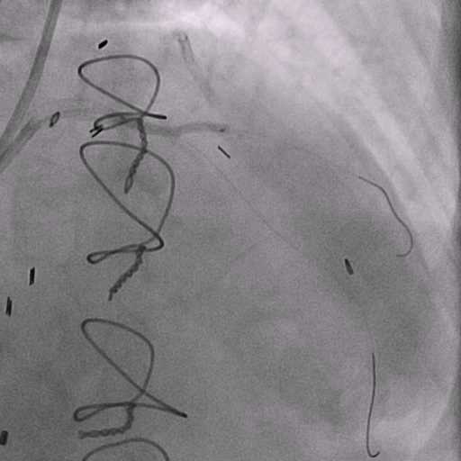
Left coronary artery angiography
- total occlusion of the left circumflex (LCx) coronary artery.
1 of 12
Left coronary artery angiography total occlusion of the left…
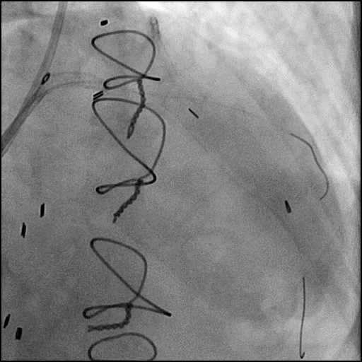
2 of 12
Left coronary artery angiography significant stenoses in the proximal…
3 of 12
LIMA to LAD bypass graft angiography.
4 of 12
Pre-dilatation of the D1 lesion with an Emerge 2.0/20mm…
5 of 12
Pre-dilatation of the proximal LAD lesion with a Trek…
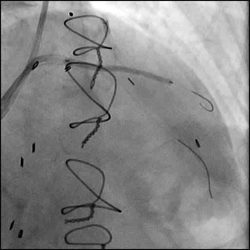
6 of 12
Angiography of the LAD-D1 after lesion pre-dilatation showing compromised…
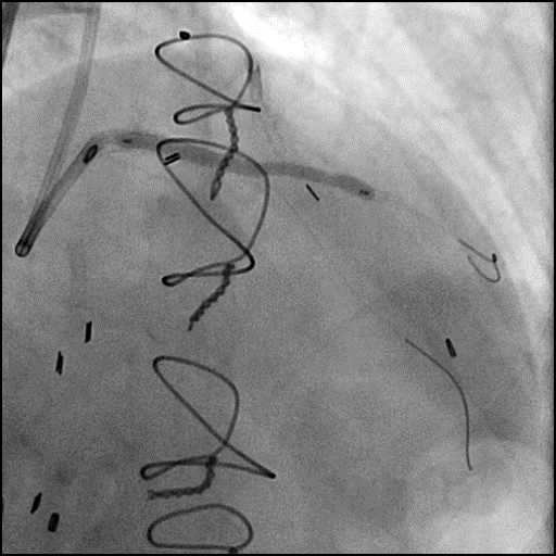
7 of 12
Deployment of a Xience Xpedition 2.75/38mm stent in the…
8 of 12
Post-dilatation of the stent placed in the LAD extending…

9 of 12
Follow up angiography of the LAD-D1 after stent post-dilatation…
10 of 12
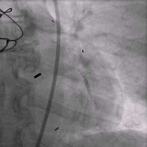
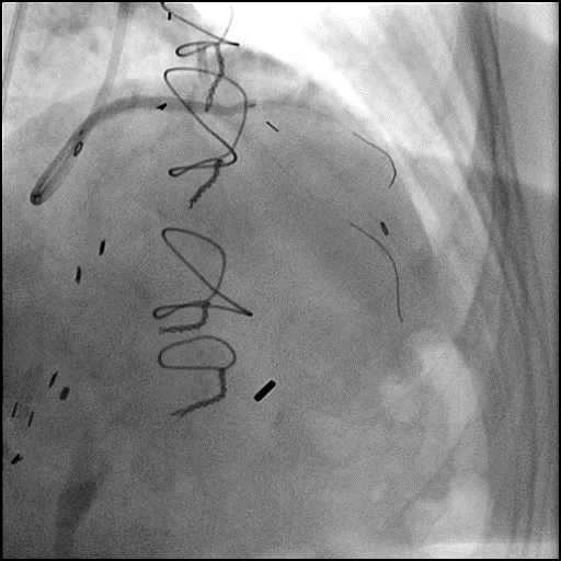
Balloon inflation to tamponade the vessel.
11 of 12
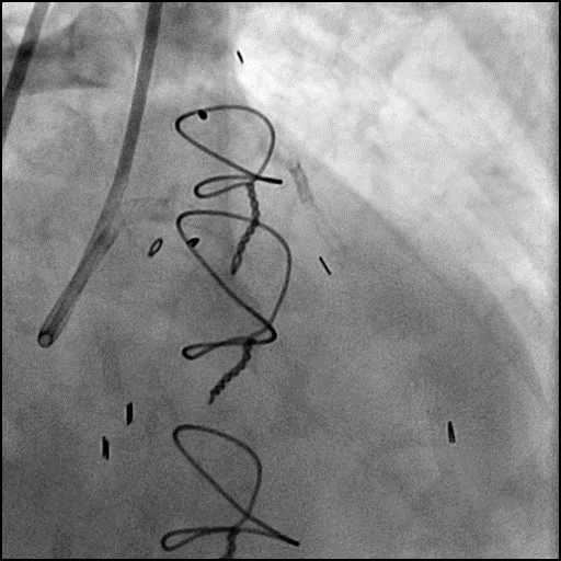
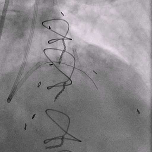
Angiography after unsuccessful attempt to deliver a covered stent…
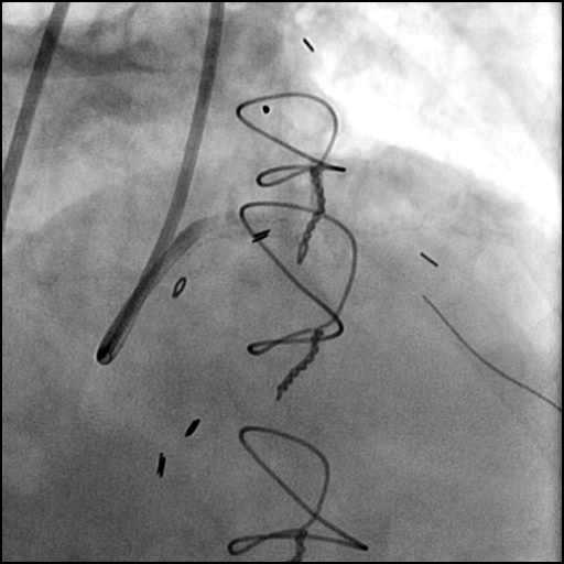
12 of 12
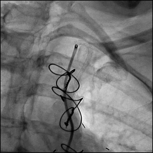
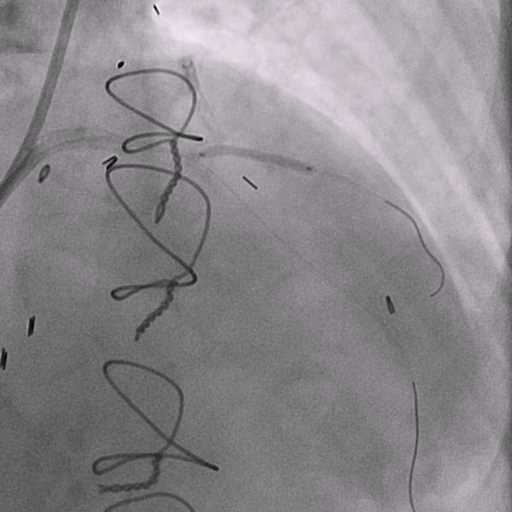
Aspiration thrombectomy was performed without improvement in flow. Angiography…
Post-procedure EKG

Post-procedure Echocardiography
Case Overview
- Underwent intervention of the proximal LAD-D1.
- Procedure was complicated by a type 3 perforation following post-dilatation of the stent.
- As per protocol, anticoagulation was stopped.
- Perforation was inadequately sealed with prolonged balloon tamponade of the vessel.
- Placement of a covered stent was attempted but unsuccessful.
- Angiography revealed D1 abrupt vessel closure due to acute thrombosis.
- Aspiration thrombectomy was performed without improvement in flow.
- Follow up angiography showed extension of thrombosis with AVC now at the level of the LM and aspiration thrombectomy was repeated without improvement in flow.
- Patient became hemodynamically unstable with on going chest pain.
- An IABP was placed, for hemodynamic support and further intervention was deferred.
- Fortunately, the patient had a history of a 3 vessel CABG with patent grafts (LIMA-LAD, SVG-LCx, and SVG-RCA).
- Echocardiography showed no evidence for pericardial effusion.
- Post procedure Troponin-I peaked at >50.0 ng/mL and CK-MB peaked at 254.4 ng/mL.
- Hospital course was further complicated by development of high grade AV block, and a PPM was placed. Patient was discharged home 10 days later.
Learning Objectives
- What is the likely explanation or reason why the complication occurred?
- Aggressive post-dilatation with a stent balloon.
- How could the complication have been prevented?
- Perform post-dilatation using a dedicated non-compliant balloon instead of using the same stent balloon. High pressure inflation of a stent balloon is unpredictable and can lead to dog-boning effect, edge dissection, perforation and other related complications. Inflation of a noncompliant balloon is true to a manufactures measurements and prevents dog-boning.
- Is there an alternate strategy that could have been used to manage the complication?
- Ellis Type 1 and 2 perforations usually seal spontaneously and are conservatively managed. Such patients should be closely monitored in the catheterization lab, and serial echocardiography should be performed, particularly if there is an Ellis Type 2 coronary perforation because it may lead to cardiac tamponade. Ellis Type 3 perforations are associated with increased risk of cardiac tamponade and mortality, and require immediate intervention/treatment. Ellis Type 3 Cavity Spilling perforation management is unclear. Usually they are conservatively managed, unless there is significant extravasation or the patient is symptomatic.
- Coronary perforation management algorithm:
- 1st: Prolonged balloon inflation: Position the balloon (or stent-balloon post stent deployment) just proximal or at the level of the perforation to prevent ongoing extravasation and development of hemo-pericardium. Ideally, the balloon to artery ratio should be 1:1. Inflate for 5-10 minutes followed by test deflations with contrast given in between inflations to evaluate the status of the perforation. If there is ongoing extravasation, re-inflate the balloon to stop further extravasation of blood into the pericardial space. This strategy helps stabilize the patients and gain control of the situation, while the operator prepares for echocardiography, pericardiocentesis, and more definitive treatment to seal the perforation.
- 2nd: Anticoagulation management: ‘STOP’ all anticoagulation immediately if you suspect or visualize a perforation. We consider ‘REVERSING’ heparin with protamine sulfate (to achieve ACT <225s) after coronary equipment is removed to prevent thrombosis within the vessel. If using bivalirudin, it can take up to 1-2 hours for its anticoagulation effect to a normalize after it is stopped. If patient was on glycoprotein IIB/IIIA inhibitors: For abciximab, consider giving platelet transfusion; tirofiban and eptifibatide have a short half life and their reversal can typically be achieved by stopping there infusion or in extreme cases with hemodialysis. Cangrelor has a short half life and its reversal can be achieved by stopping its infusion.
- 3rd: Covered stent: Standard of care for a perforation located in the proximal to mid segment of a vessel of appropriate size (≥2.5 mm), with no major side branch across the region where the stent will be placed. If a covered stent can be delivered to a distal vessel perforation, and the vessel is of appropriate size, covered stent placement to seal the perforation is reasonable. If the clinical situation allows, proceed with direct stent placement whenever possible using a single catheter or two-catheter (Ping-Pong) strategy. The stent should be quickly positioned and immediately deployed to high pressure. This should be followed by high pressure post-dilatation (18-20 atm) to achieve appropriate stent apposition.
- 4th: Embolization of distal vessel perforations: Non-surgical techniques for distal vessel embolization include: Coils, Gel Foams, Glues, Microspheres, Thrombin injection, Subcutaneous tissue, Autologous Blood Clots and multiple other agents (depending on what is available in an individual catheterization lab). Embolization leads to loss of vessel flow beyond point where embolized material is delivered and subsequent infarct in the vessel territory.
- 5th: Surgery Intervention: Ligation or suturing of the vessel for hemostasis with bypass grafting to the distal vessel. Pericardial patch/Teflon with possible bypass grafting to the distal vessel (consider this approach if vessel has multiple stents and/or presence of a subepicardial hematoma).
- Could have used a more deliverable covered stent (i.e. Papyrus Biotronik but this was not available when this case was performed).
- What are the important learning points?
- Techniques associated with perforations: use of hydrophilic/extra stiff wires, atherectomy devices, cutting balloons, increase balloon-to-artery ratio, high pressure post-dilatation of stent.
- Angiographic findings associated with perforations: CTO, severe calcification, type C lesions, eccentric lesions, RCA or LCx lesion, tortuous vessels
- Device related perforations tend to be more catastrophic than wire related perforations. This is because devices cause trauma and disrupt the integrity of the vessel wall.
- Delivery of a covered stent:
- A covered stent can be delivered using the same guide catheter after removal and retrieval of the balloon, if there is no significant hemodynamic decompromise and in the absence of a large perforation present. If using this strategy, an operator needs to act quickly because once the balloon is deflated, there will be ongoing coronary extravasation into the pericardial space.
- Alternatively, a second guide catheter strategy can be used for delivering a covered stent. To do this, obtain alternate access, advance a second guide catheter, disengage the first guide catheter and intubate the perforated artery with the new guide catheter (PING-PONG technique). Next, advance a second guidewire to the proximal edge of the inflated balloon, deflate the balloon, advance the wire to the distal vessel and then immediately re-inflate the balloon. The covered stent is advanced over the second guidewire until proximal to the inflated balloon. Then deflate the balloon and remove it along with the first guidewire (into the initial guide catheter), and quickly position the covered stent and immediately deploy it to high pressure. This should be followed by high pressure post-dilatation (18-20 atm) to achieve appropriate stent apposition.
- Despite several measures we were unable to treat the perforation as intended. Moreover, the procedure was complicated by AVC of the LM due to thrombosis. Fortunately, the patient had a 3 vessel CABG with a patent grafts (LIMA-LAD, SVG-LCx, and SVG-RCA). Therefore, further intervention was deferred after placement of an IABP.
- It is believed, patients with history of cardiac surgery (i.e CABG) may not necessarily develop cardiac tamponade because of persistence of pericardiotomy and pericardial adhesions. However, cardiac tamponade can still occur in patient with cardiac surgery due to development of a loculated effusion or repair of the pericardium at the time of surgery.
- It is important to monitor for development of a hemothorax with serial Hgb/Hct in patients with prior history of CABG when a an extravasating perforation occurs.