


Perforation Type 3 Wire – Case 4
Clinical Presentation
- 64-year-old male who presented with chest pain (CCS Class III).
Past Medical History
- HTN, HLD, DM, Former Tobacco Use
Clinical Variables
- Stress MPI: Mild to moderate ischemia with reversibility in the RCA and LCx distribution.
- Cardiac CTA: Hemodynamically significant stenosis in the LAD and RCA territories.
Medications Heading
- Home Medications: Aspirin, Rosuvastatin, Metformin
- Adjunct Pharmacotherapy: Clopidogrel, Bivalirudin
Pre-procedure EKG Heading

Angiograms
Previous
Next
1 of 11
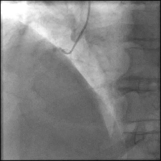
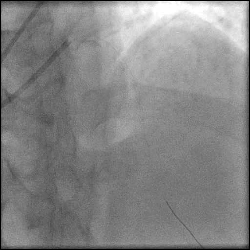
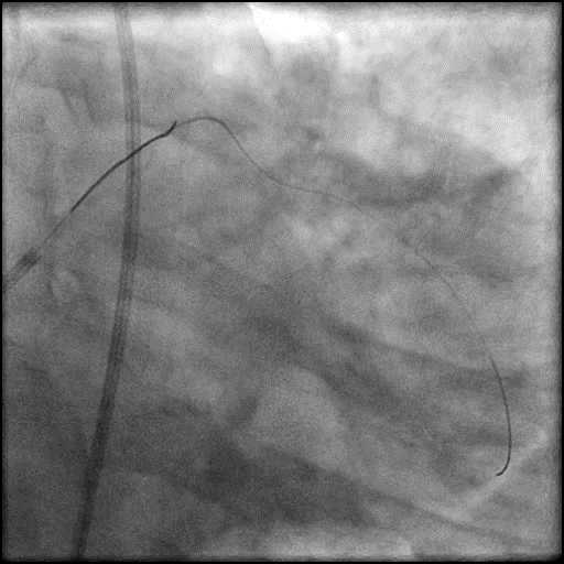
Right coronary artery (RCA) angiography
- calcified total occlusion filling via collaterals from the left circumflex (LCx) coronary artery (next video).
1 of 11
Right coronary artery (RCA) angiography calcified total occlusion filling…
2 of 11
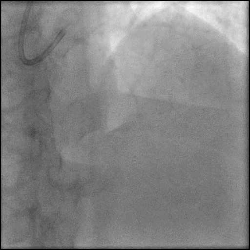
Left coronary artery angiography 80-90% severely calcified lesion in…
3 of 11
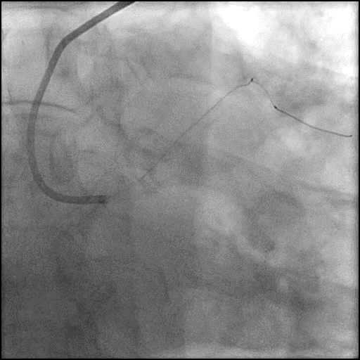
Wiring of the LAD with a rota-wire with use…
4 of 11
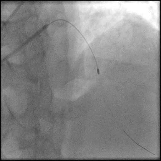
Rotational atherectomy of the LAD using a 1.5mm burr…
5 of 11
Angiography of the LAD after rotational atherectomy.
6 of 11
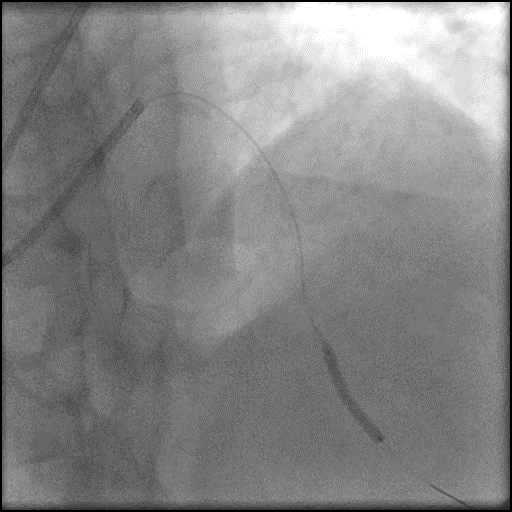
Pre-dilatation of the LAD lesion with a Trek NC…
7 of 11
Angiography of the LAD after lesion pre-dilatation.
8 of 11
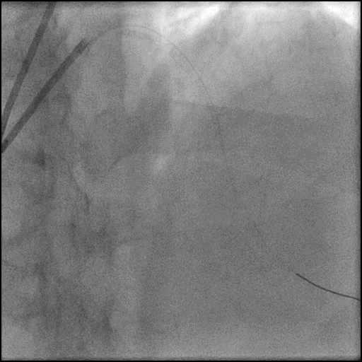
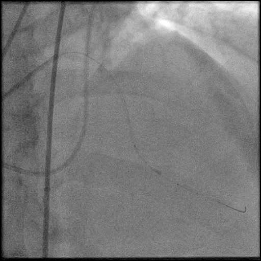
Distal wire position was lost due to patient movement.…
9 of 11
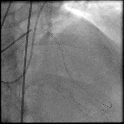
Multiple wires were used to re-cross the LAD with…
10 of 11
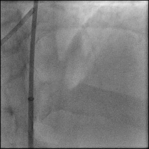
Patient developed cardiac tamponade and an emergent echocardiography guided…
11 of 11
Angiography of the LAD after serial balloon inflations showing…
Post-procedure EKG

Case Overview
- Underwent intervention of the LAD.
- IABP emergently was placed with angiography showed presence of a Type D dissection followed by AVC of the LAD.
- Patient moved during the procedure, and distal wire position was lost.
- Multiple wires were used to traverse the vessel with the procedure being further complicated by a type 3 mid vessel perforation with contrast extravasation into the pericardial space.
- Perforation was inadequately sealed with prolonged balloon tamponade of the vessel.
- Patient developed cardiac tamponade and an emergent pericardiocentesis was performed followed by placement of a pericardial drain.
- Decision was made to abort the procedure and the patient was referred for emergent CABG.
- S/p 2 vessel CABG performed with LIMA-LAD, and SVG-PDA.
- Post operative course was uneventful and he was discharged home 7 days later without any further sequelae.
Learning Objectives
- What is the likely explanation or reason why the complication occurred?
- Type 3 perforation likely wire related (multiple wires used to recross dissected segment after wire position was lost).
- Dissection likely due to aggressive balloon inflation vs. wire related.
- AVC due to progression of dissection.
- How could the complication have been prevented?
- Keep the wire in sight at all times to prevent loss of wire.
- Is there an alternate strategy that could have been used to manage the complication?
- Ellis Type 1 and 2 perforations usually seal spontaneously and are conservatively managed. Such patients should be closely monitored in the catheterization lab, and serial echocardiography should be performed, particularly if there is an Ellis Type 2 coronary perforation because it may lead to cardiac tamponade. Ellis Type 3 perforations are associated with increased risk of cardiac tamponade and mortality, and require immediate intervention/treatment. Ellis Type 3 Cavity Spilling perforation management is unclear. Usually they are conservatively managed, unless there is significant extravasation or the patient is symptomatic.
- Coronary perforation management algorithm:
- 1st: Prolonged balloon inflation: Position the balloon (or stent-balloon post stent deployment) just proximal or at the level of the perforation to prevent ongoing extravasation and development of hemo-pericardium. Ideally, the balloon to artery ratio should be 1:1. Inflate for 5-10 minutes followed by test deflations with contrast given in between inflations to evaluate the status of the perforation. If there is ongoing extravasation, re-inflate the balloon to stop further extravasation of blood into the pericardial space. This strategy helps stabilize the patients and gain control of the situation, while the operator prepares for echocardiography, pericardiocentesis, and more definitive treatment to seal the perforation.
- 2nd: Anticoagulation management: ‘STOP’ all anticoagulation immediately if you suspect or visualize a perforation. We consider ‘REVERSING’ heparin with protamine sulfate (to achieve ACT <225s) after coronary equipment is removed to prevent thrombosis within the vessel. If using bivalirudin, it can take up to 1-2 hours for its anticoagulation effect to a normalize after it is stopped. If patient was on glycoprotein IIB/IIIA inhibitors: For abciximab, consider giving platelet transfusion; tirofiban and eptifibatide have a short half life and their reversal can typically be achieved by stopping there infusion or in extreme cases with hemodialysis. Cangrelor has a short half life and its reversal can be achieved by stopping its infusion.
- 3rd: Covered stent: Standard of care for a perforation located in the proximal to mid segment of a vessel of appropriate size (≥2.5 mm), with no major side branch across the region where the stent will be placed. If a covered stent can be delivered to a distal vessel perforation, and the vessel is of appropriate size, covered stent placement to seal the perforation is reasonable. If the clinical situation allows, proceed with direct stent placement whenever possible using a single catheter or two-catheter (Ping-Pong) strategy. The stent should be quickly positioned and immediately deployed to high pressure. This should be followed by high pressure post-dilatation (18-20 atm) to achieve appropriate stent apposition.
- 4th: Embolization of distal vessel perforations: Non-surgical techniques for distal vessel embolization include: Coils, Gel Foams, Glues, Microspheres, Thrombin injection, Subcutaneous tissue, Autologous Blood Clots and multiple other agents (depending on what is available in an individual catheterization lab). Embolization leads to loss of vessel flow beyond point where embolized material is delivered and subsequent infarct in the vessel territory.
- 5th: Surgery Intervention: Ligation or suturing of the vessel for hemostasis with bypass grafting to the distal vessel. Pericardial patch/Teflon with possible bypass grafting to the distal vessel (consider this approach if vessel has multiple stents and/or presence of a subepicardial hematoma).
- What are the important learning points?
- Avoid performing complex procedures at a facility where surgical backup is not immediately available as a rescue option.
- Placement of a pericardial drain prior to transferring a patient with a type 3 perforation and cardiac tamponade is essential.
- When shifting the patient from the cardiac catheterization lab to the operating room, it is vital to continuously aspirate the pericardial drain in a sterile manner if there is a persistent symptomatic effusion/cardiac tamponade.