


Slow Flow – Case 1
Clinical Presentation
- 86-year-old male who presented with chest pain (CCS Class III).
Past Medical History
- HTN, HLD, DM, Former Tobacco Use, CAD s/p 3 vessel CABG (SVG to RCA known to be occluded) followed by multiple PCI’s, Prostate Cancer s/p Brachytherapy (2011), OSA, Aortic Stenosis (AVA 0.8 cm2), ESRD on iHD
- LVEF 37%
Clinical Variables
- None
Medications
- Home Medications: Aspirin, Clopidogrel, Atorvasatin, Metoprolol Tartrate, Isosorbide Mononitrate, Gabapentin, Cyclobenzaprine, Allopurinol, Phoslo, Insulin
- Adjunct Pharmacotherapy: Clopidogrel, Bivalirudin, Heparin IV
Pre-procedure EKG

Angiograms
Previous
Next
1 of 25
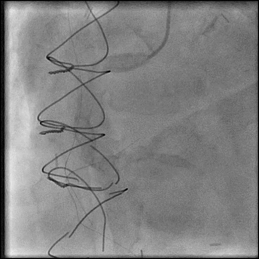
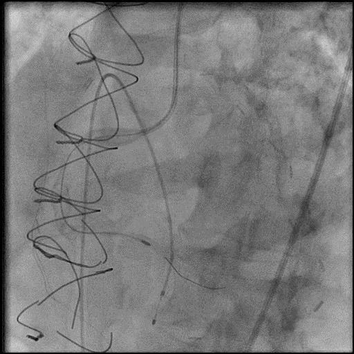
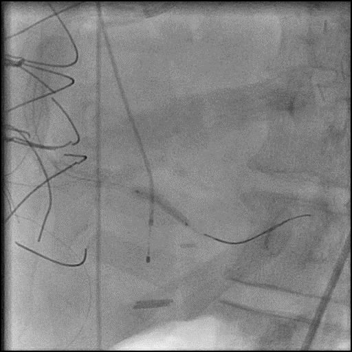
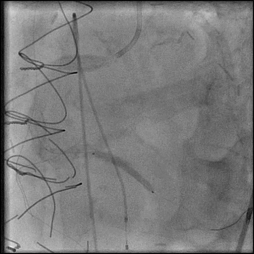
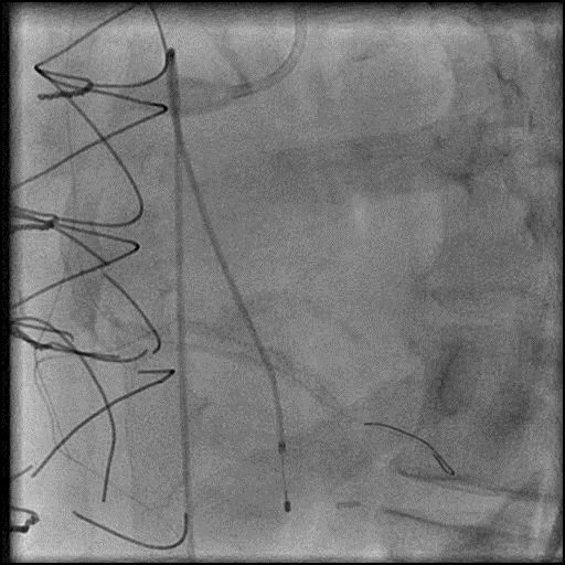
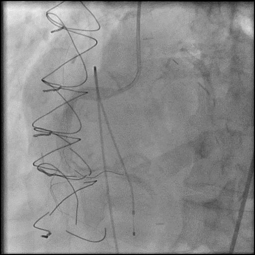
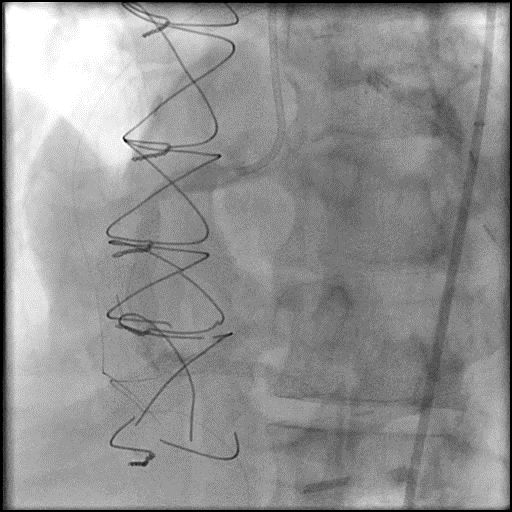
Right coronary artery (RCA) angiography
- 80-90% severely calcified ectatic lesions in the tortuous RCA
- 90-95% calcified lesion in the right posterior descending artery (RPDA).
1 of 25
Right coronary artery (RCA) angiography 80-90% severely calcified ectatic…
2 of 25
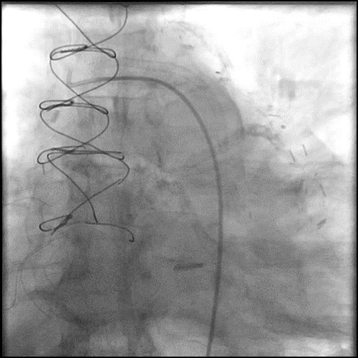
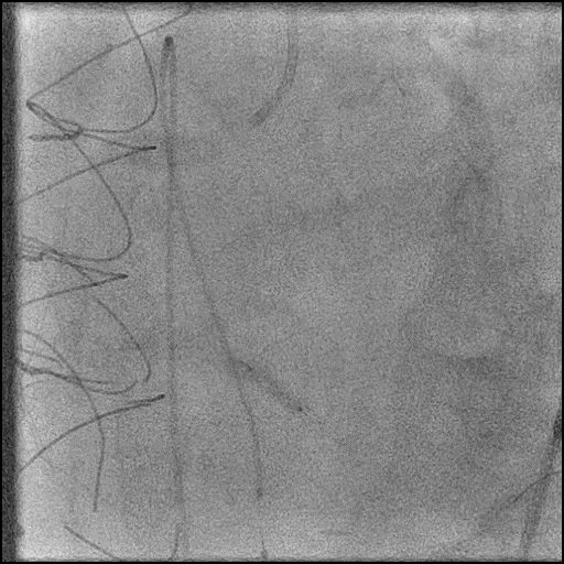
No significant stenosis in the saphenous vein graft (SVG)…
3 of 25
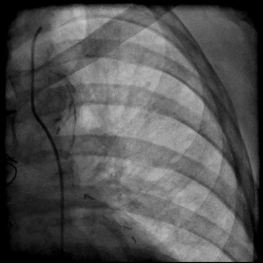
No obstruction in LIMA to LAD bypass graft.
4 of 25
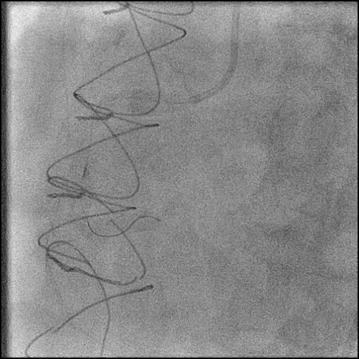
RPDA wired with use of a Teleport microcatheter.
5 of 25
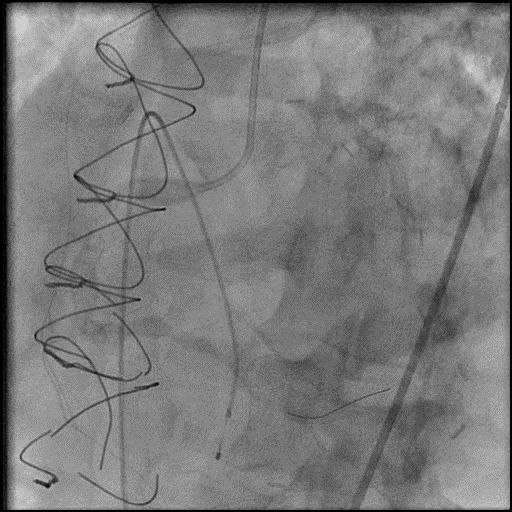
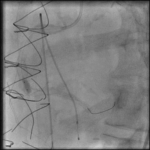
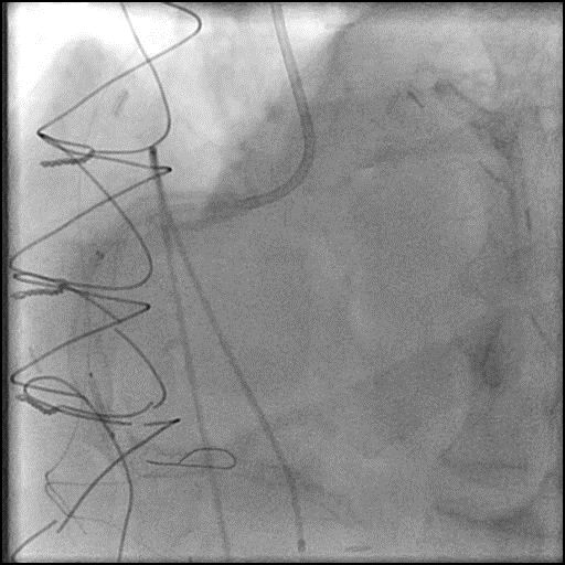
Wire exchanged for Rota-Extra Support wire and rotational atherectomy…
6 of 25
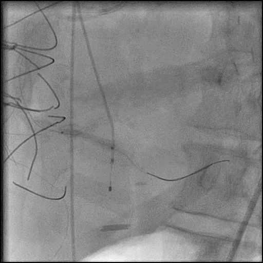
Angiography after rotational atherectomy.
7 of 25
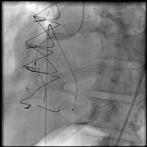
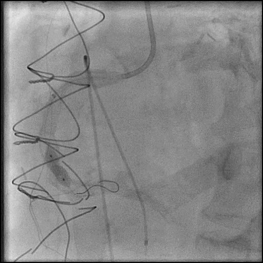
Repeat rotational atherectomy using a 1.75mm burr at 150k…
8 of 25
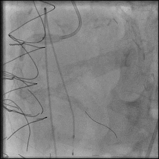
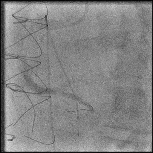
Angiography after rotational atherectomy showing slow flow.
9 of 25
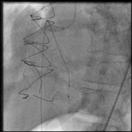
IC vasodilators administered through the guide catheter with no…
10 of 25
Pre-dilatation of the distal RCA with a Trek NC…
11 of 25
Guidezilla extension catheter used to help deliver a NC…
12 of 25
Pre-dilatation of the RPDA with a NC Emerge 2.75/8mm…
13 of 25
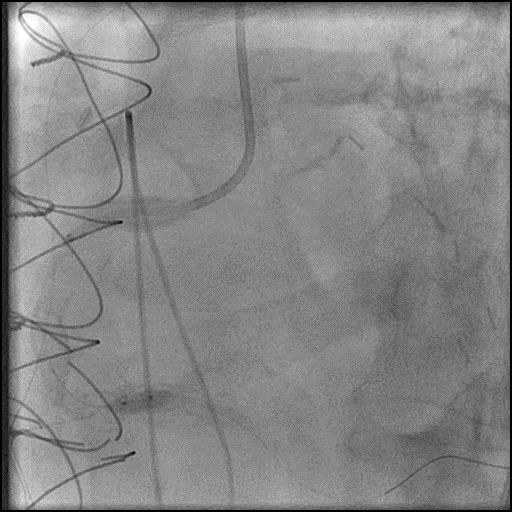
Deployment of a Synergy 2.75/38mm stent in the RPDA.
14 of 25
Post-dilatation of the stent placed in the RPDA with…
15 of 25
Pre-dilatation of the proximal RCA with a Trek NC…
16 of 25
Pre-dilatation of the proximal RCA with a Trek NC…
17 of 25
Angiography of the RCA after proximal RCA lesion pre-dilatation.
18 of 25
Pre-dilatation of the RCA (distal to proximal) with a…
19 of 25
Deployment of a Synergy 5.0/28mm stent in the distal…
20 of 25
Deployment of a Synergy 5.0/12mm stent in the proximal…
21 of 25
Angiography of the RCA after placement of two additional…
22 of 25
Positioning of a Synergy 5.0/12mm stent in the mid…
23 of 25
Post-dilatation of stents placed in the RCA with a…
24 of 25
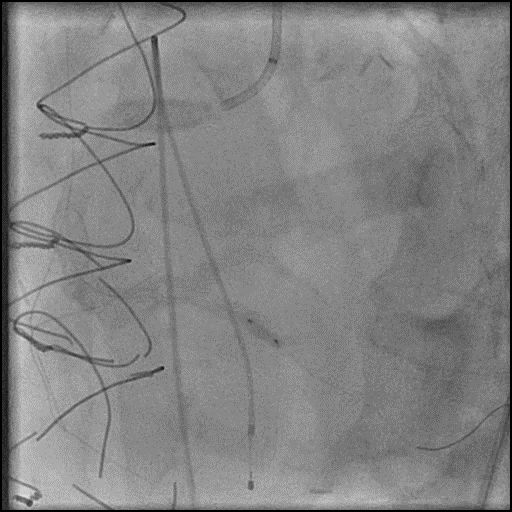
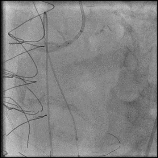
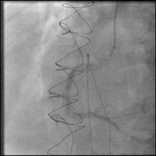
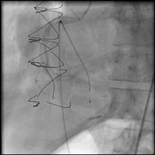
Angiography after stents post-dilatation showing persistent slow flow.
25 of 25
Final angiography showing TIMI 2 flow in the RCA.…
Post-procedure EKG

Case Overview
- Underwent intervention of the RCA.
- Rotational atherectomy was performed, and after upsizing to a 1.75mm burr the procedure was complicated by slow flow.
- IC vasodilators were administered through the guide catheter, without improvement in flow.
- Serial balloon inflations performed followed by placement of multiple stents in the RCA, without improvement in flow.
- Patient was hemodynamically unstable with ongoing chest pain, and an IABP was placed.
- Troponin-I peaked at 32.06 ng/mL and CK-MB peaked at 73.8 ng/mL.
- Patient was discharged 4 days later without further sequelae.
Learning Objectives
- What is the likely explanation or reason why the complication occurred?
- Slow flow following rotational atherectomy – likely due to distal embolization of debris.
- How could the complication have been prevented?
- When performing rotational atherectomy consider the following to prevent complications:
- Keep systolic BP >100mmHg and avoid bradycardia
- Perform short runs <20 seconds with a gentle pecking motion to advance the rota burr, maintain rotational speed around 150-160k RPM
- Use Rota-flush and consider use of glycoprotein IIB/IIIA inhibitors
- Vasodilators should be given prophylactically and for treatment of slow flow/no-reflow.
- Assure patient is given adequate periprocedural antithrombotic therapy (antiplatelets and anticoagulants).
- Pay close attention to the ACT during the procedure and dose anticoagulation accordingly to maintain ACT >300 prior to performing an intervention (Hemochron machine).
- Use a rota-burr which is appropriately sized for the vessel. Ideally the burr should be 0.5-0.6x the distal vessel size when performing a plaque modification strategy, and 0.8-0.85x the distal vessel size when performing a debulking strategy (STRATA trial). With plaque modification strategy the purpose is to disrupt the calcium so it allows for safe passage of devices and easier expansion of device, balloons, and stents. With debulking, the aim is to break the calcium into particles so it can move through the coronary artery and eventually washout of the coronary microcirculation.
- Lithotripsy device: Currently under investigation in the USA (approved for use in Europe). It is uncertain if this device will result in reduced complications including slow flow.
- Is there an alternate strategy that could have been used to manage the complication?
- The initial step in management of slow flow/no-reflow involves administration of intra-coronary vasodilators through the guide catheter. If this fails, recommended using a dual-lumen microcatheter (Twinpass is the only dual lumen microcatheter available in the USA) to deliver intra-coronary medications to the distal vessel and microvasculature. Next, perform angiography with delivery of contrast through the microcatheter to determine if there is distal coronary flow. If distal vessel flow is not preserved the likely etiology of abrupt vessel closure (AVC ) is no-reflow due to distal embolization of debris or thrombus, and IC vasodilators should be administered through the microcatheter targeted to the distal vessel and microvasculature. If flow is preserved, then the likely etiology of AVC is dissection (proximal to the point of microcatheter injection), and treatment involves placement of a stent. It is reasonable to perform aspiration thrombectomy prior to microcatheter based angiography injection (depending on the clinical context/presence of thrombus).
- What are the important learning points?
- The best treatment for slow flow/no-reflow is to prevent it from happening.
- The exact mechanism of the no-reflow phenomenon is unclear, but it is thought to be associated with endothelial swelling, neutrophil infiltration, and platelet aggregation causing obstruction and spasm in the microvasculature.
- One of the possible complications of PCI especially during rotational or orbital atherectomy is slow flow/no-reflow. The common settings where this complication arises are when performing interventions of long calcified lesions, CTO’s (particularly RCA CTO), thrombotic lesions, and vein grafts. Also, incidence is higher when a patient has poor LV function and hemodynamic instability.
- Important to have multiple vasodilators readily available during a procedure. We use the following agents and administer them intra-coronary.
- Nitroprusside 50-200 mcg, Adenosine 30-40 mcg, Verapamil 100-200 mcg, Nicardipine 100-200 mcg
- Nitroglycerin 100-200 mcg (we use NTG for slow flow/no-reflow when it involved the epicardial vessels and not the coronary microvasculature)
- If the patient is hypotensive and this impedes the administration of intra-coronary vasodilators to treat slow flow/no-reflow, we recommend administration of IV phenylephrine 100-200 mcg as needed (may result in reflex bradycardia) to increase blood pressure, and then administer intra-coronary vasodilators.
- If there is refractory slow flow/no reflow then consider placement of an IABP. This helps with reduction in afterload, and improves coronary perfusion pressure by increasing coronary blood flow during diastole, and reduction in LVEDP.
- Depending on the size and function of the LV (LVEF <30%), consider using upfront LV support as it can help improve coronary perfusion pressure.