Case 14: Calcified LM/LAD lesion treated with RA and lithotripsy
Case Presentation
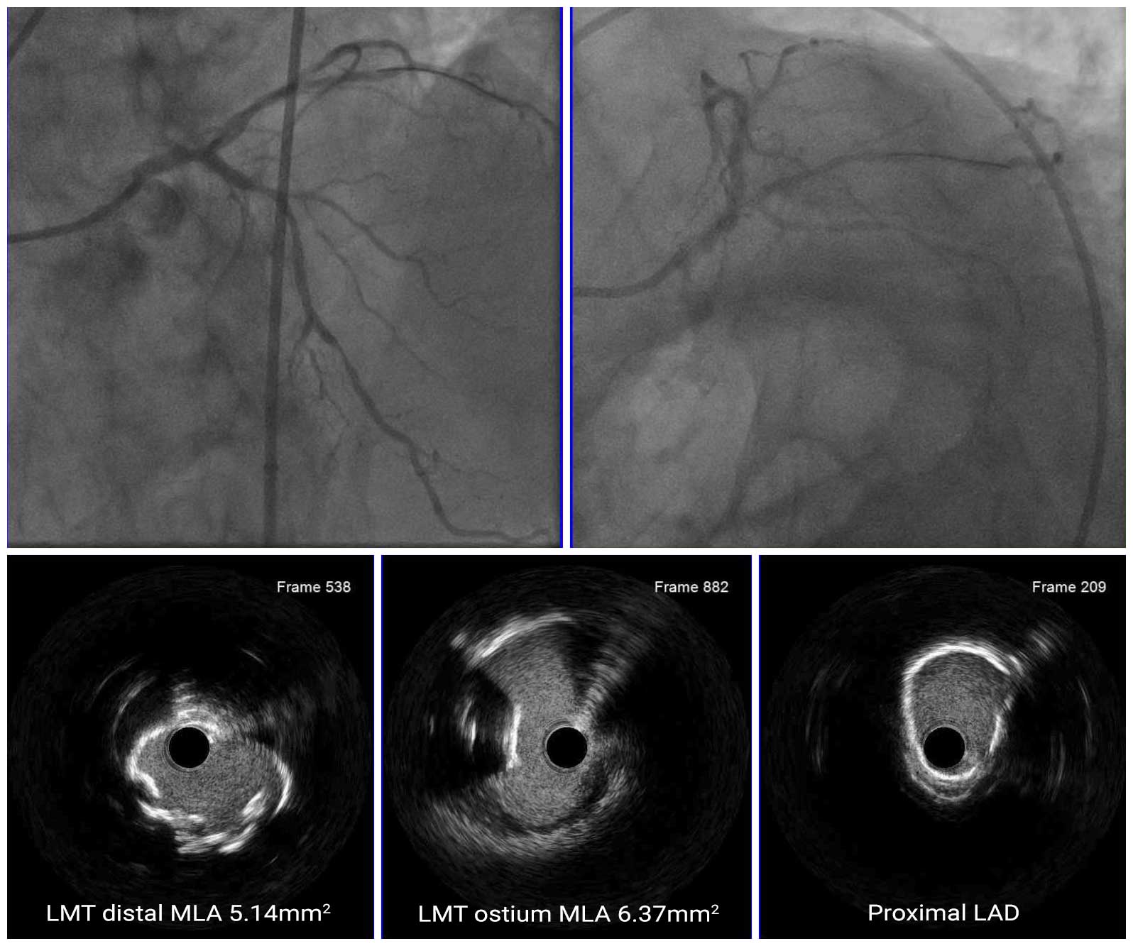
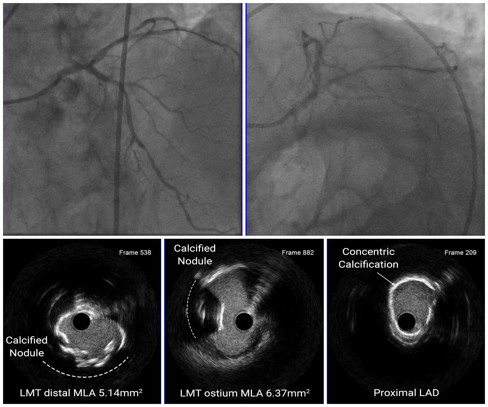
A 70-year-old male had presented with angina on exertion for 1 week to outside hospital. He had associated exacerbation of CHF and found to have LM disease, and transferred to our hospital for staged PCI. He had a history of prior MI, hypertension, and hyperlipidemia. Echocardiography showed globally reduced left ventricular wall motion, particularly in the apex wall. Coronary angiography showed 80-90% stenosis in the distal LAD, 70-80% stenosis in the proximal LAD, and 60-70% stenosis in the distal LM.
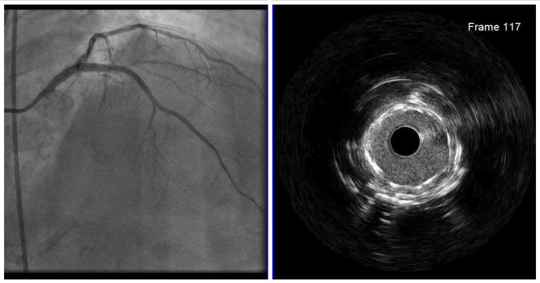
Angio Pre
IVUS Pre
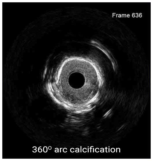
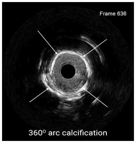
IVUS pullback showed a circumferential calcified lesion in the proximal LAD, a circumferential calcified lesion with calcified nodule in the distal LM, and a calcified nodule in the ostial LM. The MLA was 5.14mm2 in the distal LM and 6.37mm2 in the ostial LM.
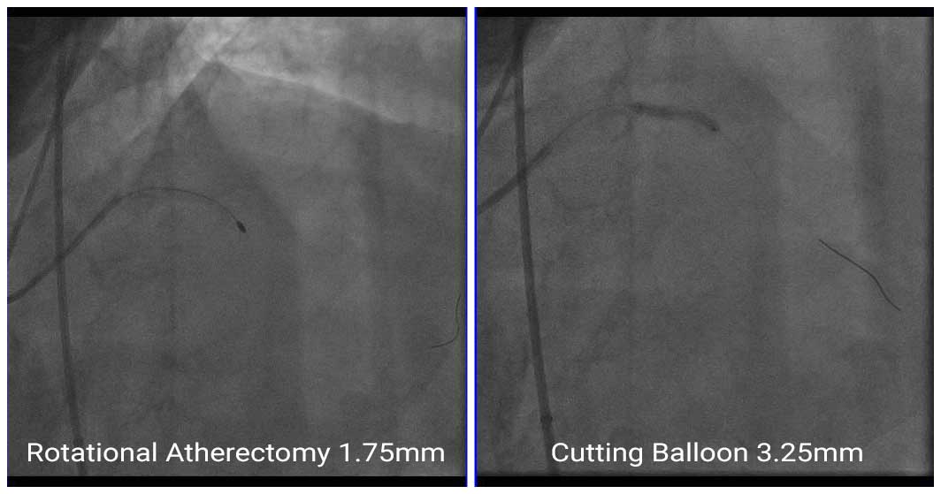
Since the proximal LAD lesion was highly calcified, rotational atherectomy (RA) with a 1.75mm burr was performed. Subsequently, a 2.5mm x 12mm DES was deployed in the lesion at the distal LAD. As for the proximal LAD lesion, pre-dilatation was performed with a 3.25mm cutting balloon up to 6atm, but adequate dilation was not achieved.
IVUS After RA
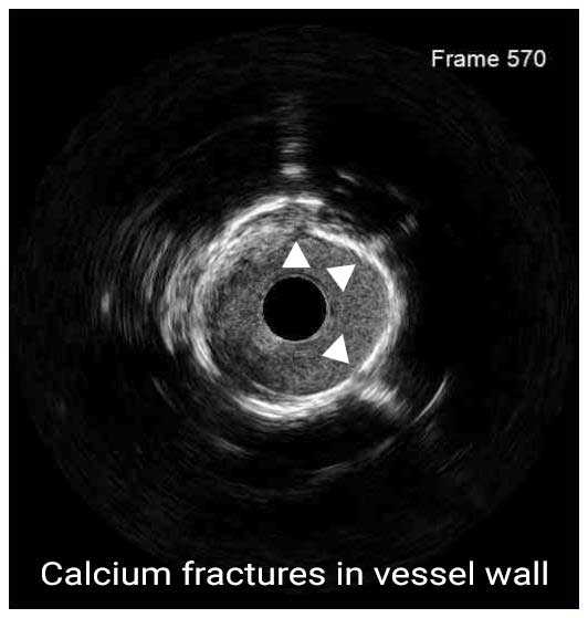
IVUS After IVL
A 3.5mm x 24mm DES was deployed at the proximal LAD and a second 4.0mm x 28mm DES was deployed at the proximal LAD to the LM ostium, with both stents overlapping.
Angio Post
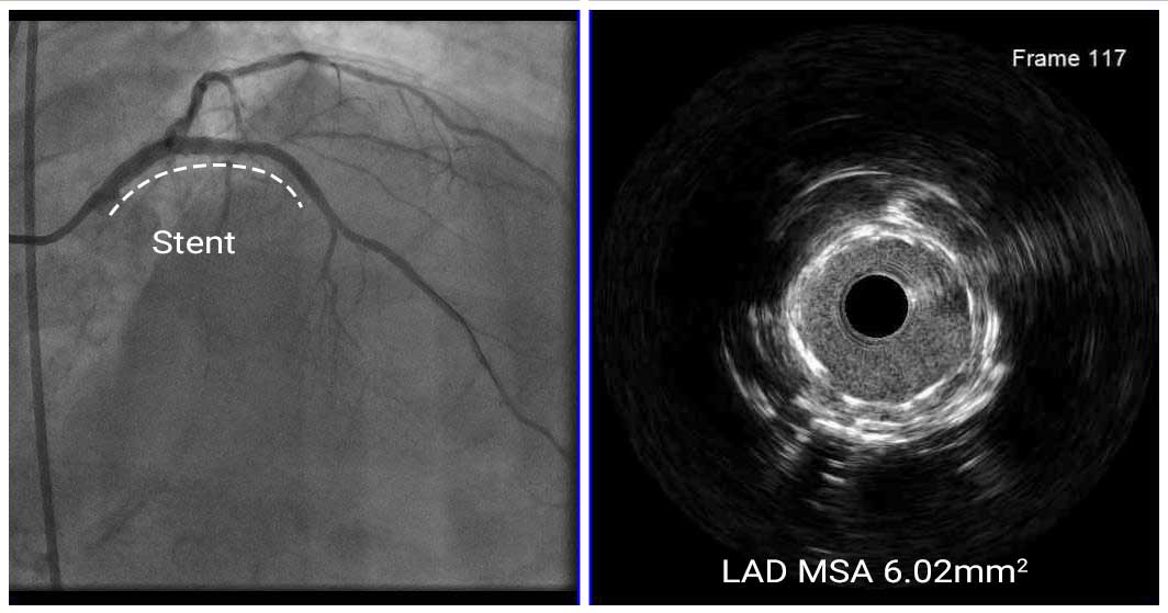
Post-Stent IVUS
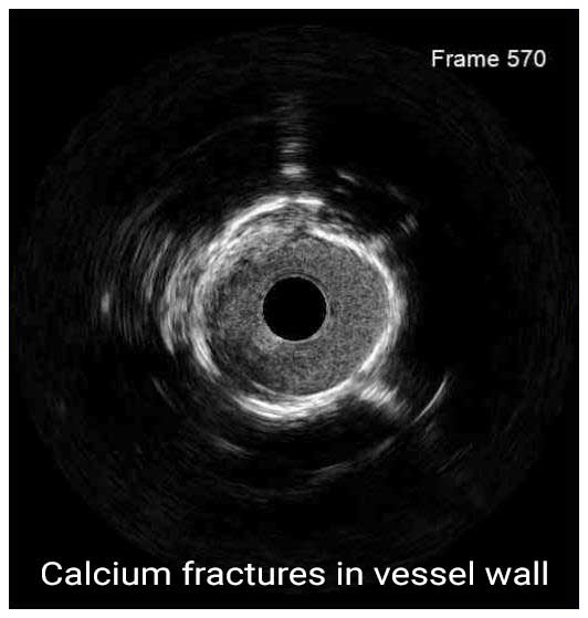
In this case, the presence of the old stent implanted in the proximal LAD could not be recognized on angiography due to heavy calcification. However, IVUS revealed that a stent had been previously implanted. We were able to perform IVL on a highly calcified lesion that could not be ablated adequately with the RA. IVUS confirmed calcium fractures, which increased the chances for adequate expansion on subsequent POBA and stent deployment.

