Case 15: Stent malapposition in ectatic LAD
Case Presentation
A 60-year-old male came from an outside hospital with chest pain as his chief complaint. Coronary angiography was performed by his previous physician which revealed a 2-vessel lesion with ostial LAD 90%, mLAD 80%, and pLCX 90%. He was admitted to our hospital for high risk PCI of LAD. He has a history of chronic diastolic heart failure, hypertension, IDDM, hyperlipidemia, ESRD (on dialysis), and COPD.
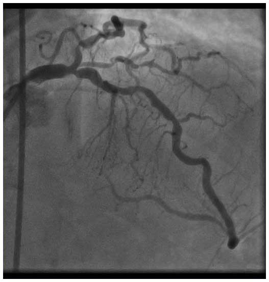
Angio Pre
Since the calcification was very severe on angiography, we first performed rotational atherectomy (RA) with a 1.75mm burr. The IVUS after RA showed an ectatic lesion with calcified plaque which could also be confirmed by angiography. The MLA was 3.8mm2, distal reference was 4.1mm x 4.2mm (Lumen), and proximal reference was 6.5mm x 7.1mm (EEL-EEL).
IVUS after RA
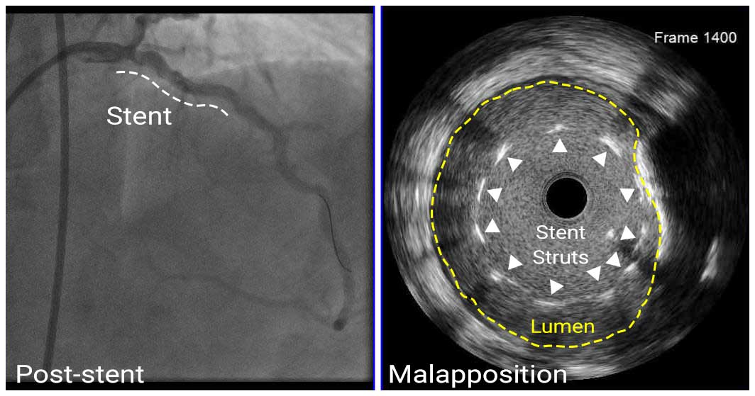
After performing pre-dilatation with 4.0mm non-compliant (NC) balloon, we implanted a DES (4.0mm x 38mm) to the lesion (pLAD-dLM). IVUS showed stent malapposition in the proximal edge of the stent due to the ectatic lesion.
Angio after stenting
IVUS post-stent
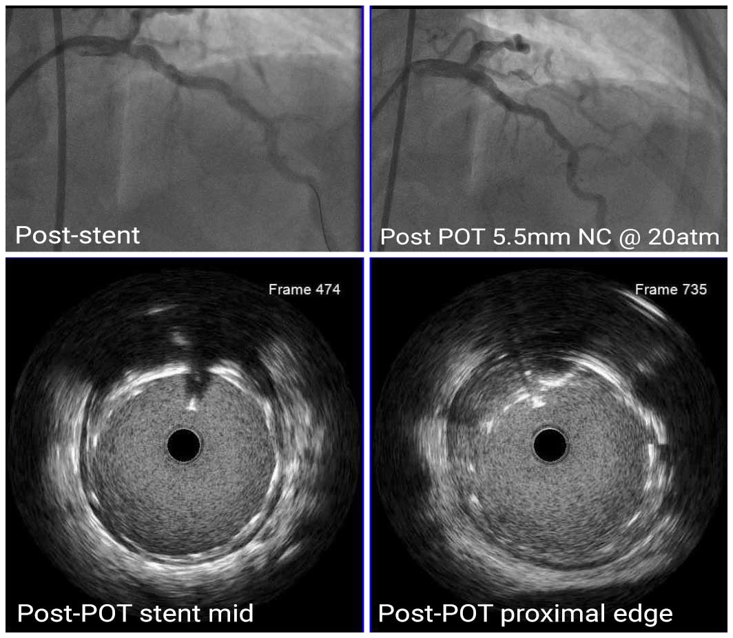
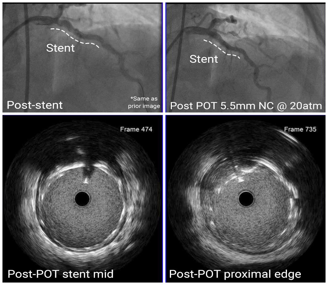
Although unrecognizable from angiography, the IVUS showed severe malapposition in the proximal part of the stent. To resolve the malapposition, we performed POT with a 5.5mm NC balloon dilated to 20 atm.
Angio Final, after POT 2x
IVUS after POT
After POT with 5.5mm NC balloon, we confirmed well-apposed stent struts with IVUS.
In this case, we were able to recognize the stent malapposition and optimize stent placement by using IVUS.


