Case 19: Distal stent edge dissection after LAD bifurcation PCI
Case Presentation
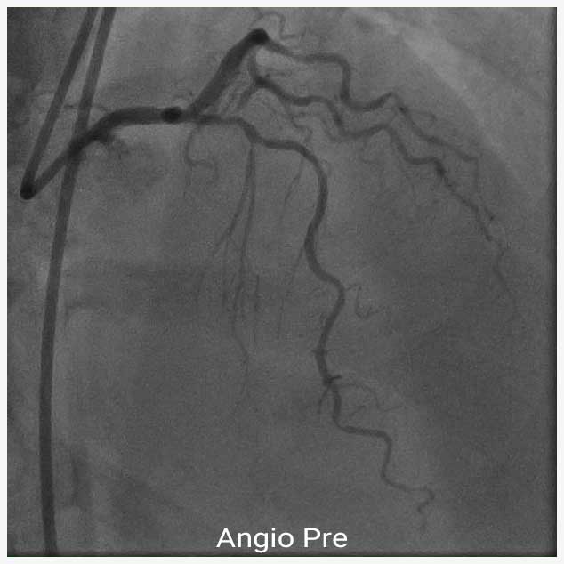
A 53-year-old female presented with CCS class III angina to our hospital. The patient had a history of hypertension, diabetes mellitus, and hyperlipidemia. CT angiography showed severe stenosis in the proximal and mid LAD. Coronary angiography revealed severely calcified bifurcation lesion with 80% stenosis in the proximal –mid LAD and a 90% stenosis in the first diagonal branch (D1).
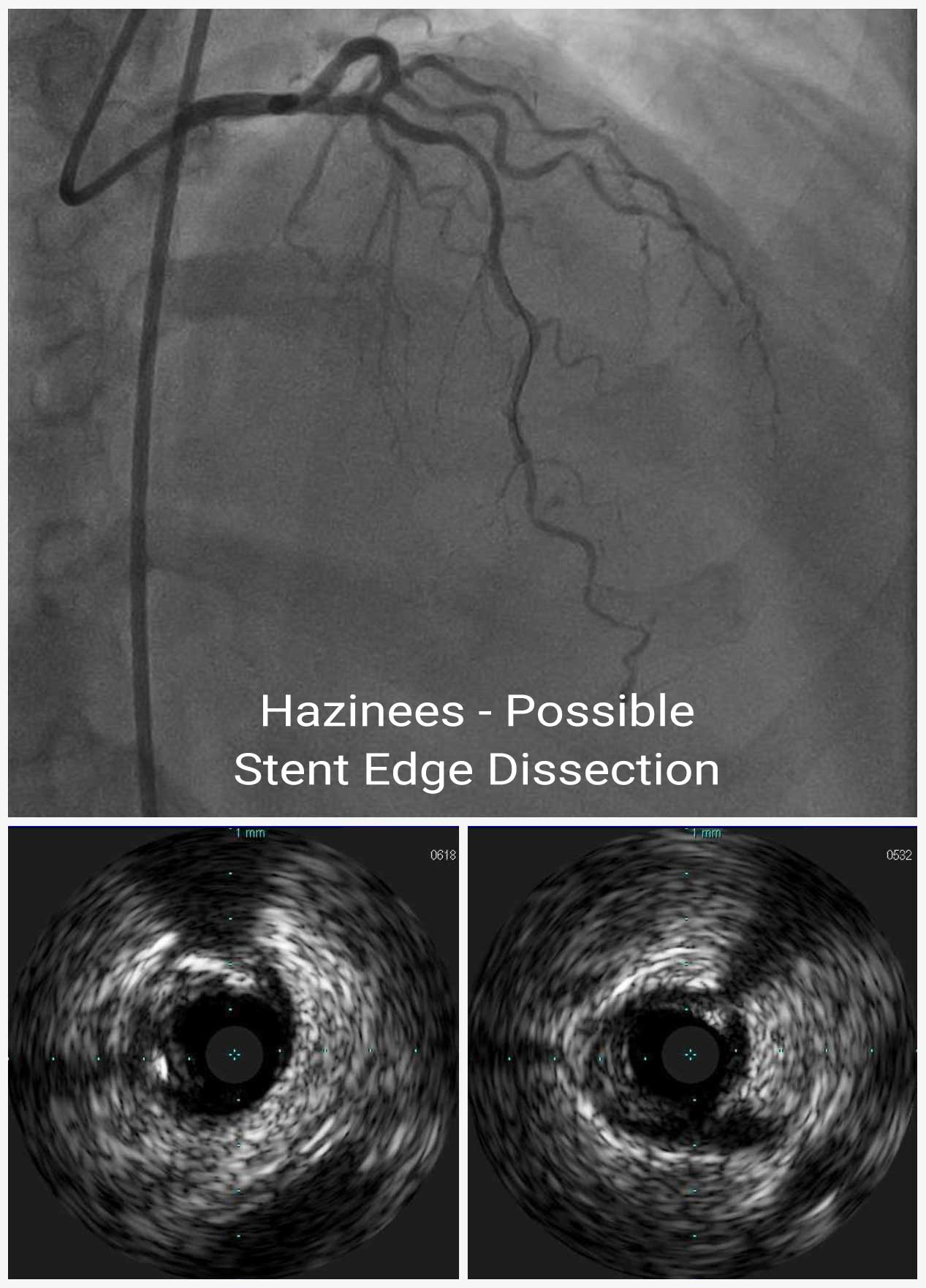
First, we performed lesion modification by using rotational atherectomy with 1.5mm burr. After pre-dilatation with non-compliant balloon in both LAD and D1, mini crush 2-stent technique was used to stent lesion with a 3.0 x 15mm and a 2.25 x 12mm DESs deployed in the LAD and D1, respectively. Post-stent angiography demonstrated TIMI 3 flow and detected slight haziness in the distal edge of the LAD stent suggesting a possibility of a distal stent edge dissection, which was confirmed by IVUS.
Post-Stent IVUS
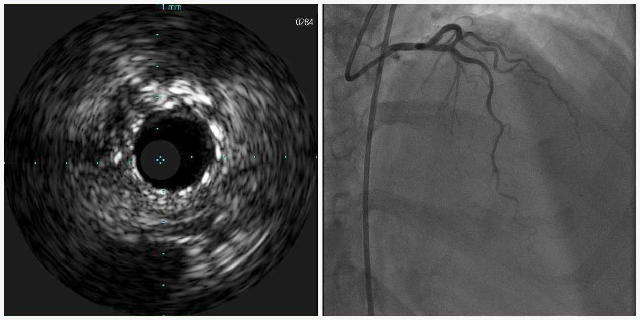
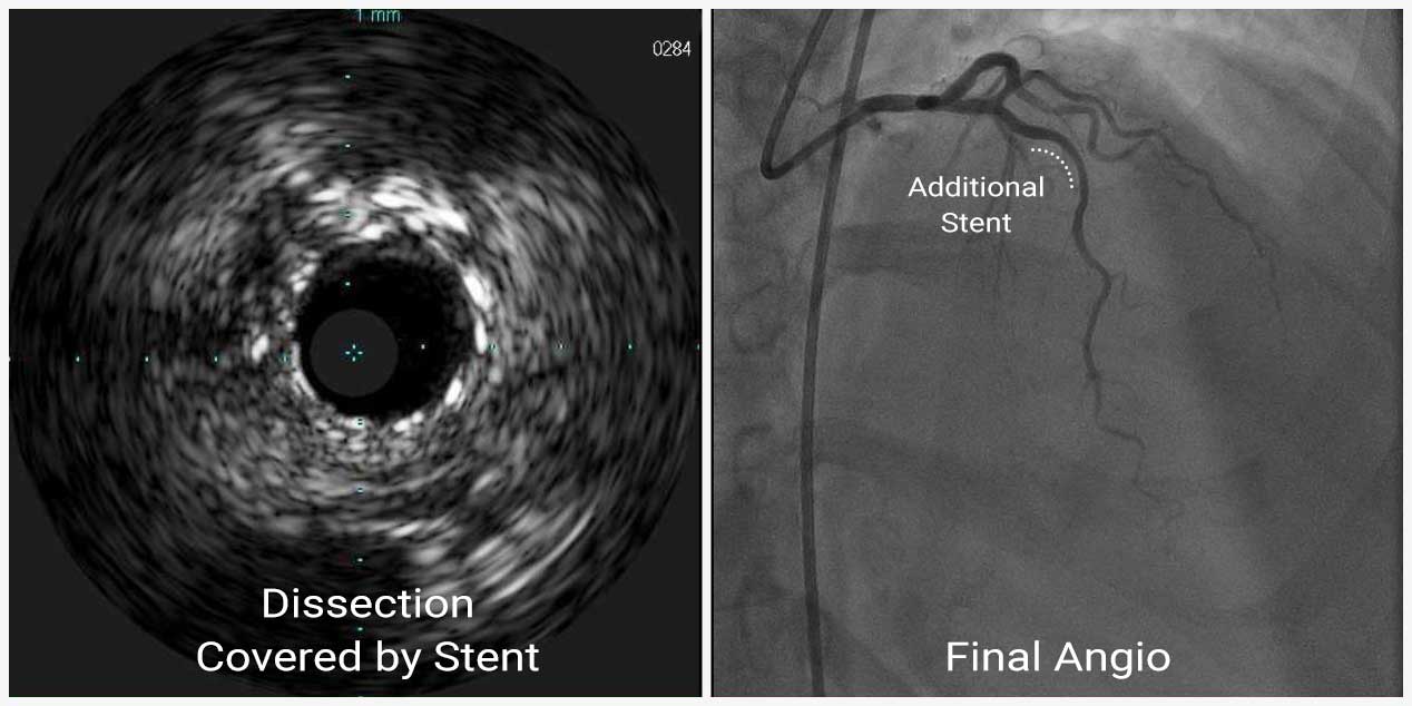
The distal stent edge dissection had two intimal flaps with a total angle greater than 60° and longitudinal length more than 2mm on IVUS, therefore, an additional 2.75 x 12mm DES was implanted into the mid LAD to cover the dissection. Final angiography and IVUS confirmed optimal post-procedural result.
Dissection covered by additional stent
In this case, we used IVUS to estimate the extent of the distal stent edge dissection, which was not clearly visualized by angiography. IVUS plays an important role in identifying and characterizing stent edge dissection, one of procedural complications after PCI. After stenting, coronary artery dissection may occur at both edges of the stent and might lead to acute coronary occlusion. Stent edge dissection is more likely to cause serious complication when it occurs at the distal edge of the stent because the dissection flap promotes lumen occlusion.

