Case 20: Combined IVUS and NIRS assessment of intermediate LAD lesion
Case Presentation
A 60-year-old male presented with CCS class II angina and positive stress test. Coronary angiography revealed a total occlusion in the LCX and a 30-50% stenosis in proximal and mid LAD. The patient had a history of hypertension, NIDDM, and hyperlipidemia. First, we performed PCI of the LCX, then we used dual-modality intravascular imaging, near infrared spectroscopy (NIRS) and IVUS, to evaluate the LAD lesion.
IVUS Pullback
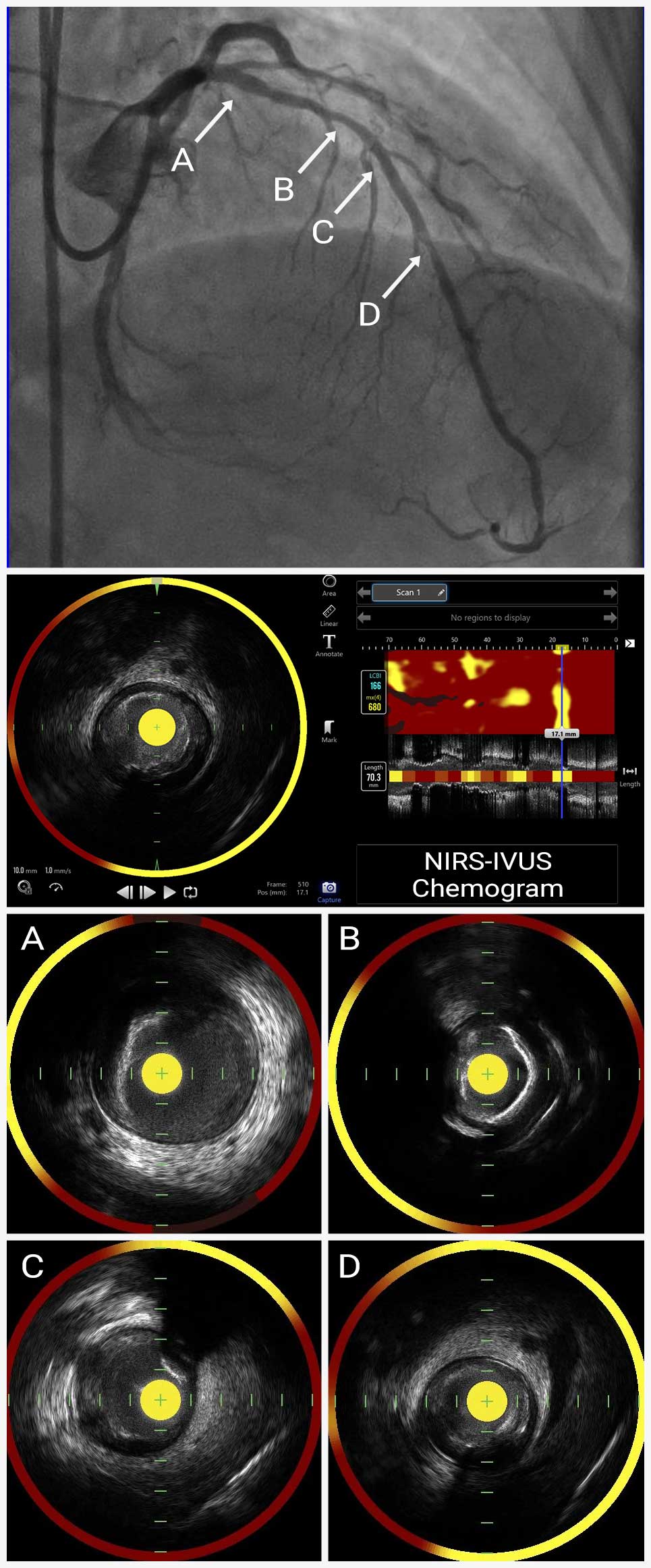
NIRS/IVUS can identify lipid-core plaques (LCP) associated with increased risk of ACS and peri-procedural complications. In addition to gray-scale IVUS cross-section, NIRS chemogram displays the distribution of lipid depending on the probability of the presence of lipid-rich plaques (LRPs). In the chemogram, areas where lipid plaque are detected are shown in yellow, areas that are unlikely to be a lipid plaque are shown in red, and areas that cannot be determined due to guidewires or other factors are shown in black. The size of lipid plaque is quantitatively assessed as lipid core burden index (LCBI) of the whole lesion or a 4mm segment (maxLCBI4mm) is calculated by dividing the number of yellow pixels by the total number of pixels available, multiplied by 1000. MaxLCBI4mm corresponds to the maximum value of LCBI calculated every 4mm within the region of interest, and its location is also displayed in the chemogram.
In this case, MLA of the LAD observed by IVUS was 3.9mm² and the maxLCBI4mm was measured 680 by NIRS suggesting high lipid content in the lesion. Based on NIRS/IVUS findings, stenting for the lesion was deferred and the patient’s lipid lowering therapy was maximized.

