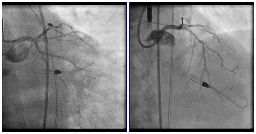
Case 8: IVUS-guided LM trifurcation PCI
Case Presentation
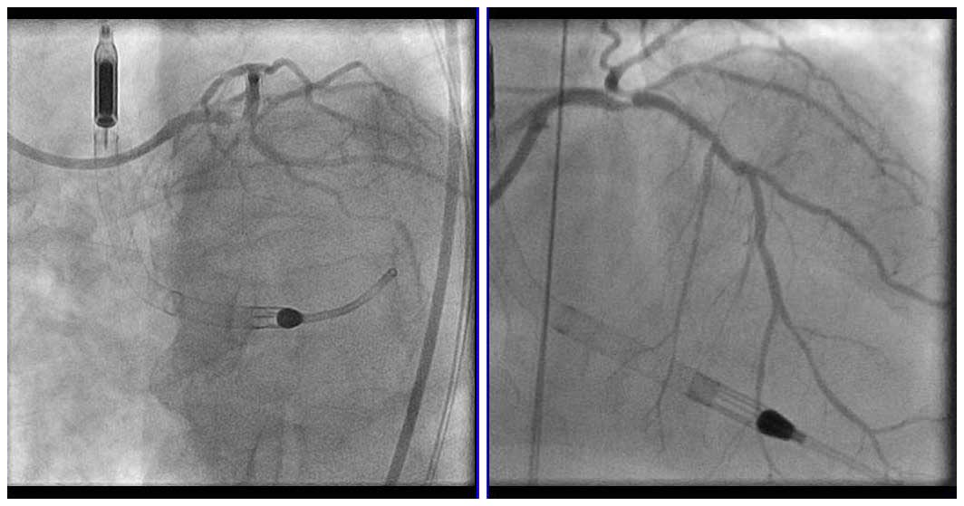
A 74-year-old male with chest pain for the past day came to outside hospital for coronary angiography, which showed 95% stenosis of the LM, 80-90% stenosis of the proximal LCX, and 80-90% stenosis in the ramus intermedius. He has a history of hypertension, NIDDM, and hyperlipidemia and has had multiple PCI procedures. The diagnosis was non-STEMI with worsening chronic heart failure, therefore the decision was made to perform Impella-assisted PCI. Echocardiography showed reduced left ventricular systolic wall motion (Ejection Fraction of 40-45%).
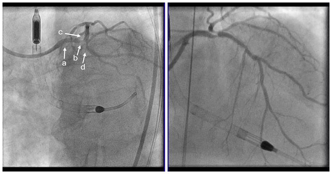
Angio Pre
Further evaluation was done with IVUS on the LAD and LCX. IVUS showed that the stenosis of the Ostium in the LAD was not severe, therefore we decided to perform provisional stenting from the proximal LCX to the LM (1 stent strategy).
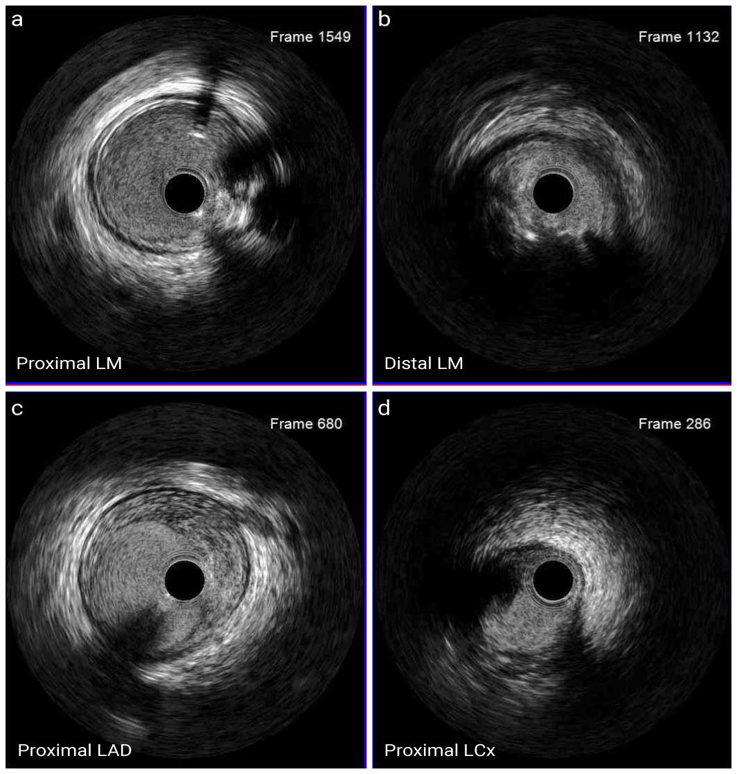
A-C; IVUS Pre LAD-LM
D; IVUS Pre LCx-LM
IVUS showed mixed plaque (fibrous, fatty, calcified) in distal LM, but no severe calcification, and fibro-fatty plaque in proximal LCX.
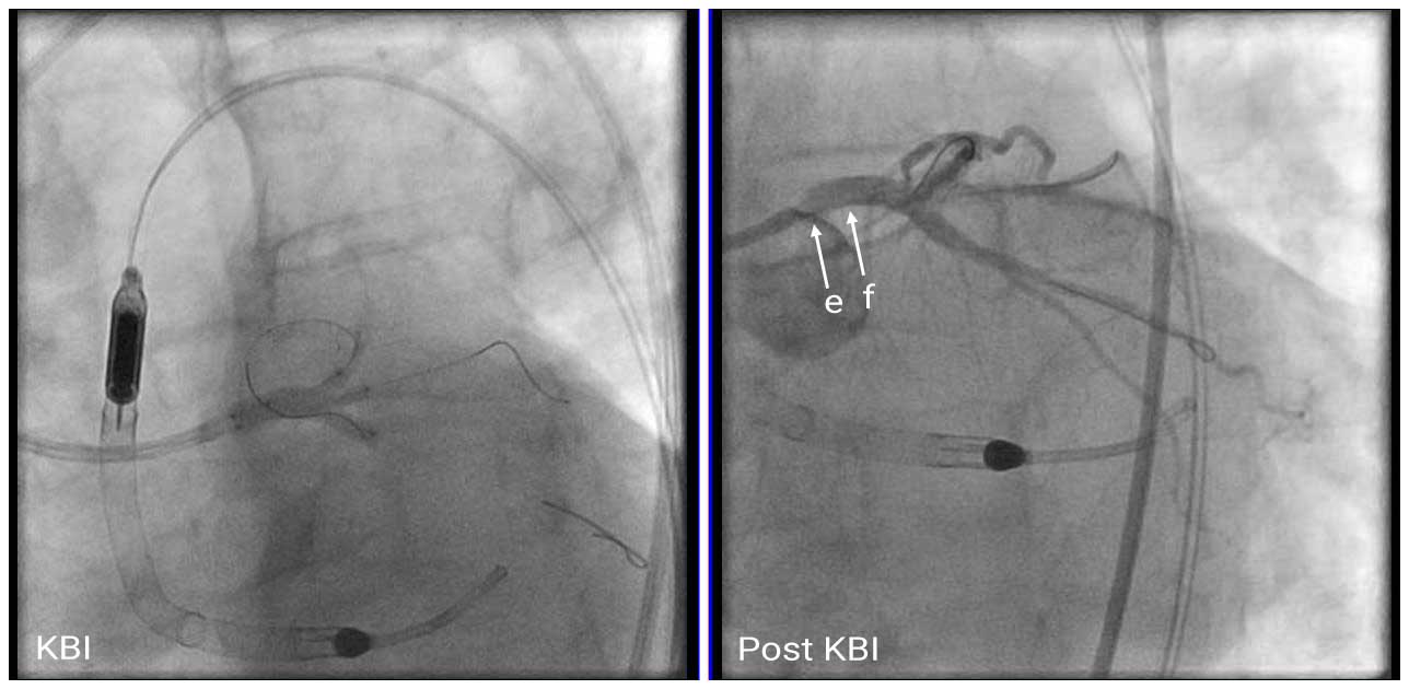
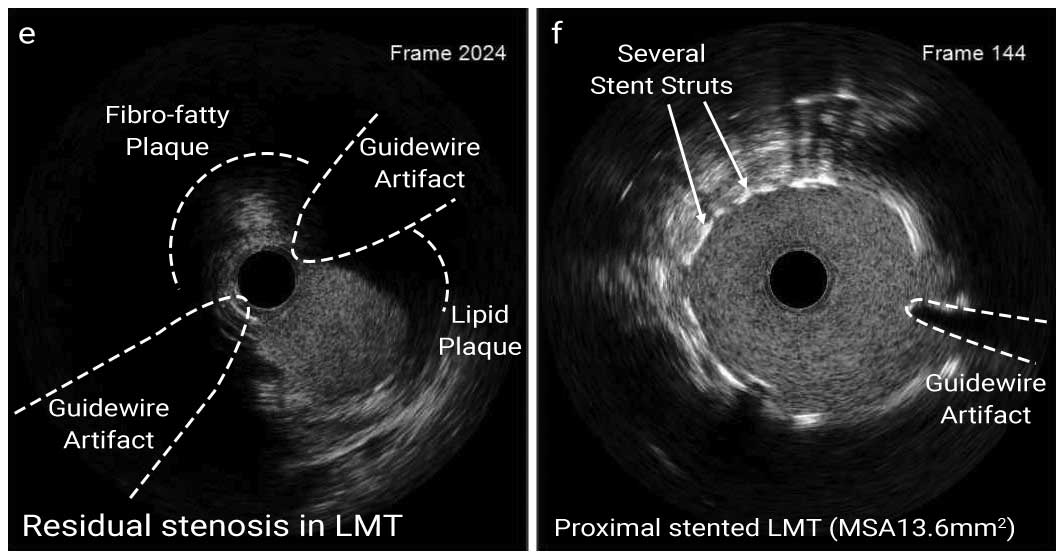
A 3.5mm x 23mm DES was deployed from distal left main to proximal LCX lesions. Serial KBI in proximal LAD/Ramus and Ramus/LCX were performed and then final KBI in proximal LAD, Ramus and proximal LCX was performed. Repeat IVUS showed residual stenotic lesion in ostial LM, therefore 4.0mm x 8mm DES was deployed in the ostial LM. The PCI was completed after confirming the absence of malapposition and dissection in the IVUS.
IVUS Proximal LM
In this trifurcation case, we performed provisional stenting in the LCX-LM direction because we confirmed the presence of the lesions at the ostial LAD and ostial LM, which could not be seen clearly by angiography. Since lesions at the ostial LM tend to be underestimated in angiography, it is important to disengage the guide catheter from the ostial LM and observe the ostium site carefully with IVUS.

