Case 9: Calcified in-stent restenosis treated with ELCA
Case Presentation
A 75-year-old male presented with new onset CCS class III angina to our hospital. Patient has history of hypertension, IDDM, hyperlipidemia and has had multiple PCI procedures. Most recently, 5 months ago, DES were implanted mid LAD (2.5mm x 38mm, 2.75mm x 18mm) at outside hospital. Echocardiography showed good left ventricular systolic function (Ejection Fraction of 65%). A coronary angiogram showed 80-90% stenosis of the mid LAD.
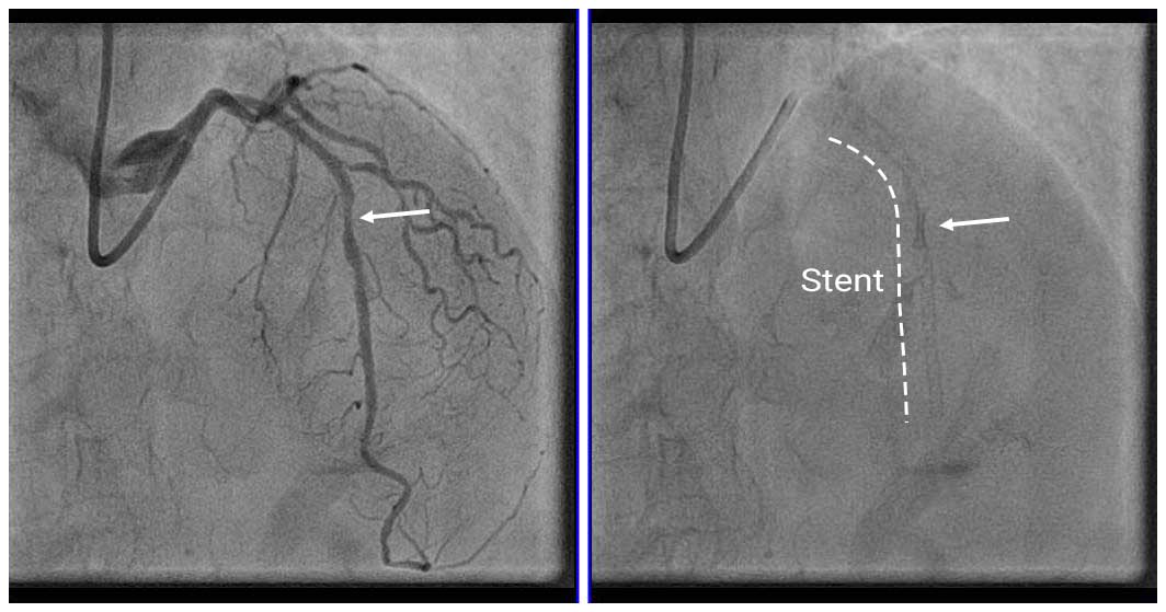
Angio Pre
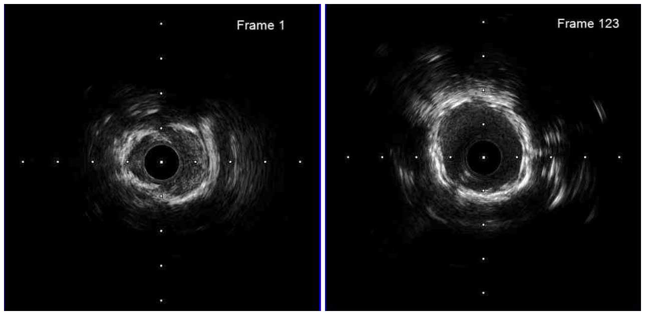
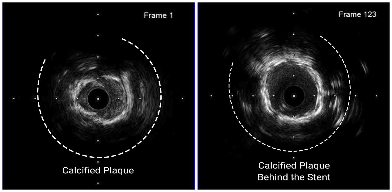
Coronary angiography showed a highly underexpanded stent that could be seen without contrast in the mid LAD. We attempted to confirm the lesion with IVUS, but it was difficult to cross the lesion due to the high degree of stenosis. As far as we could see, the stent was underexpanded and there was severe calcification outside the stent.
IVUS Pre; couldn’t cross the lesion
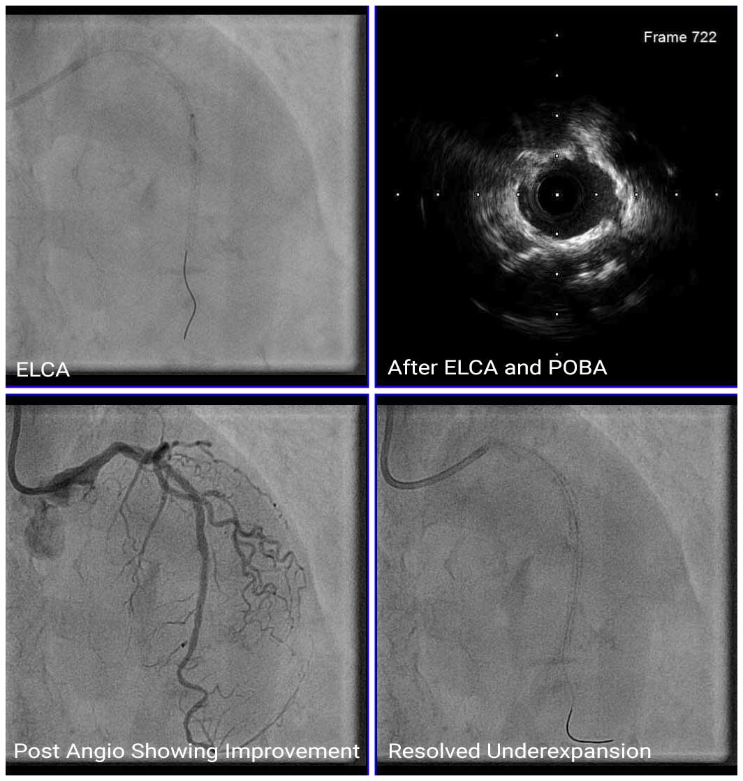
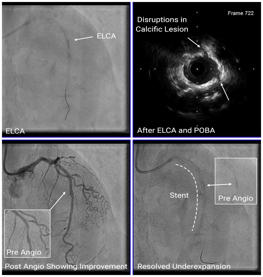
Therefore, we performed dilatation with a 2.0mm non-compliant (NC) balloon and performed ELCA with contrast medium. Then, the balloon size was increased step by step, and after dilation with a 2.5mm balloon, IVUS showed disruptions in the calcification behind the stent. Seeing the calcium modifications, we dilated the lesion with 3.5mm NC balloon up to 14 atm pressure and completed the PCI.
Angio Post
IVUS Post
Stent underexpansion is a risk factor for in-stent restenosis and stent thrombosis. In this case, after ELCA, we were able to confirm the disruptions of calcified plaque beneath the stent strut by IVUS, and additional non-compliant balloon dilatation was effective in expanding the under-expanded segment of the prior stent.


