


Acute Stent Thrombosis
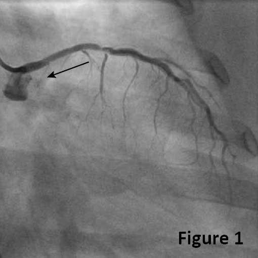
A 49-year-old female with controlled hypertension and hyperlipidemia, uncontrolled DM, multiple prior PCI, and recent NSTEMI had a PCI with a 2.5x12mm DES to the proximal LCX. Thirteen hours after the intervention, the patient developed chest pain and elevated troponin (cTnI 12.6). Coronary angiogram confirmed stent thrombosis resulting in total occlusion of the LCX (Figure 1, arrow).
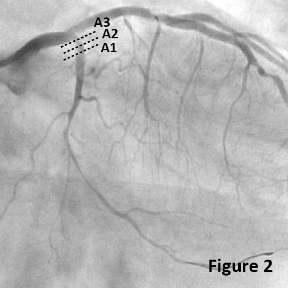
Coronary angiogram performed after thrombus aspiration and postdilatation with a 2x12mm non-compliant balloon demonstrated TIMI3 flow and a filling defect in the stent consistent with thrombus (Figure 2, A1-A3).
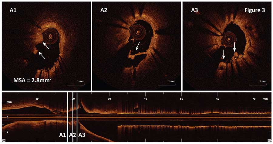
OCT imaging of the proximal LCX visualized residual thrombus inside the stent (Figure 3A1-A3, arrows) and detected stent under-expansion with the minimal stent area of 2.8mm2 (A1).
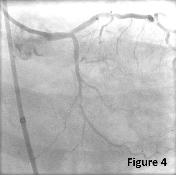
Postdilatation with 2.75×12 mm non-compliant balloon at 20atm led to satisfactory angiographic results (Figure 4). In this case, OCT helped uncover stent under-expansion as the main mechanism of acute stent thrombosis and led to the decision to proceed with postdilatation to achieve optimal stent expansion.
Ask the Experts

Ask the Experts
We are ready to help you.
ask now
ask now
Take Quiz

Take Quiz
Know about OCT? Test your knowledge.
try it now
try it now
Meet the Team

Meet the Team
Read about the team behind the apps.
learn more
learn more
Contact Us

Contact Us
Reach out to us with general comments or questions.
connect now
connect now