Combined Mechanical and Biological Etiology of DES-ISR
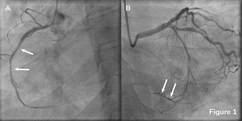
A 35-year-old male, former smoker, with a history of hypertension, hyperlipidemia and multiple PCIs of proximal-RCA, mid-RCA (unknown stent sizes), and proximal LAD (Xience Sierra 3.5x18mm) presented with anginal chest pain. SPECT MPI showed posterolateral ischemia. Coronary angiogram showed ISR lesions in the proximal and mid RCA (Figure 1A, arrows). Collateral filling of the totally occluded distal RCA can be seen in Figure 1B (arrows).
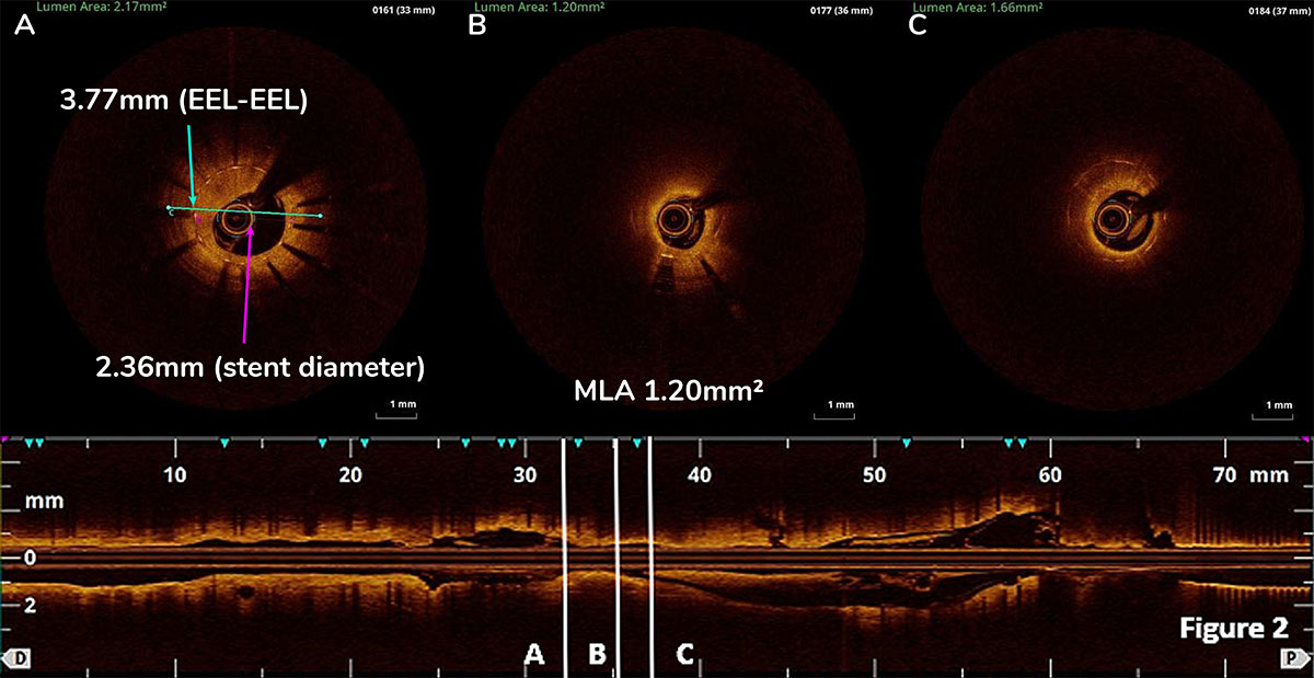
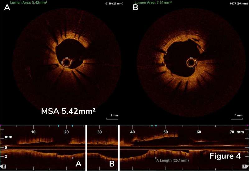
Post OCT pull back revealed plaque modification of non-calcified neoatherosclerosis with better expansion of the original stent with MSA of proximal RCA 5.42mm2 (Figure 4). In this case, OCT showed a combination of mechanical and biological etiology of DES-ISR without the presence of calcification and subsequently determined the optimal treatment option.
A composited lined up Pre and Post pullback video may help better appreciate the differences before and after intervention. Side branches were used as landmarks and marked with an apostrophe.


