Intravascular Lithotripsy for Severely Calcified Coronary Lesions
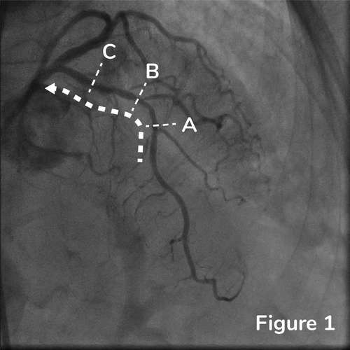
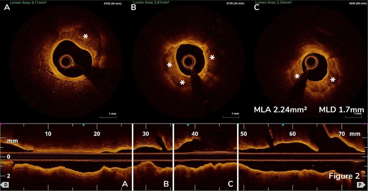
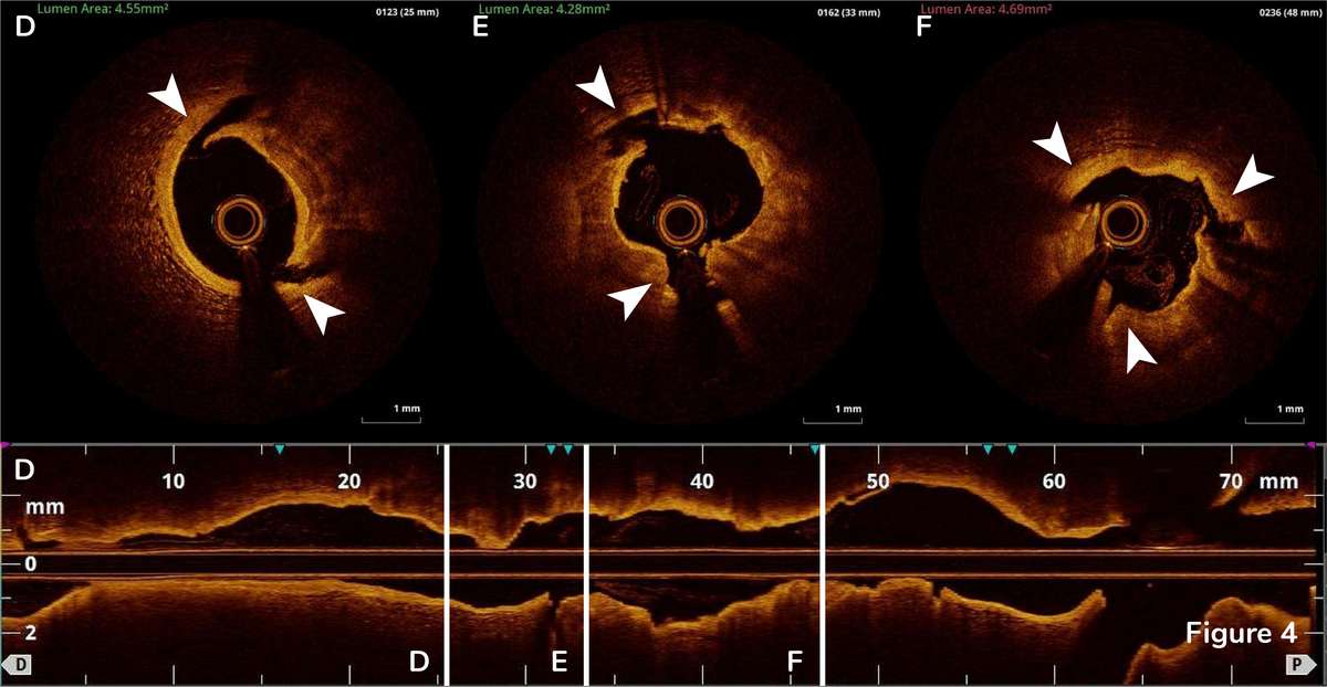
85-year-old female with coronary risk factors (diabetes mellitus, hypertension, and hyperlipidemia) presented with CCS III angina and positive SPECT MPI with anterior apical and inferior ischemia. A cardiac cath revealed 2V CAD: multiple lesions in RCA and heavily calcified 90% mid LAD lesion with a SYNTAX score of 22 and normal LV function. The patient underwent successful intervention of multiple RCA lesions with DES. The patient is now planned for staged PCI of calcified LAD PCI. Coronary angiogram showed proximal to mid LAD lesion with heavy calcification without dye injection. (Figure 1) OCT cross-sectional images correspond to their respective annotated location in angiograms throughout this case.
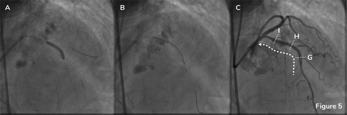
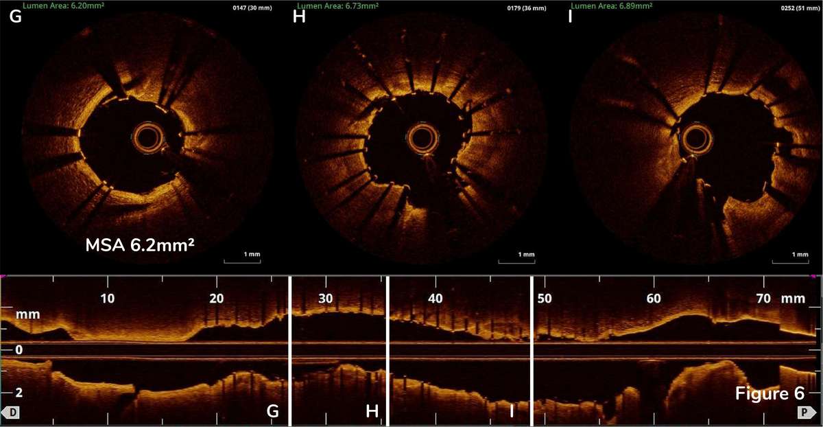
We performed 3.5x15mm high-pressure balloon dilatation with 18 atm (Figure 5A) and put 3.0x38mm DES on proximal to mid LAD (Figure 5B). Post-stenting angiography showed satisfactory results (Figure 5C). Post-stenting OCT pullback revealed a well-expanded stent with no malapposition and edge dissection (Figure 6G-I). Post-intervention MSA of LAD was 6.2mm2 (Figure 6G).
Coronary intravascular lithotripsy is a new calcium modification technology. The acoustic pressure waves from the balloon modify calcium and safely and effectively facilitates stent delivery and optimize stent expansions.1 OCT is the only device that can detect the calcium thickness and depth of calcium fracture after IVL.
Link to complete live case relay from January 29, 2022:
https://www.youtube.com/watch?v=RWr5i60VbzY
- Hill JM, Kereiakes DJ, Shlofmitz RA, Klein AJ, Riley RF, Price MJ, Herrmann HC, Bachinsky W, Waksman R, Stone GW, Disrupt CAD III Investigators. Intravascular Lithotripsy for Treatment of Severely Calcified Coronary Artery Disease. J Am Coll Cardiol 2020;76:2635-2646

