


Orbital Atherectomy in Calcified Lesion
A 71-year-old female with hypertension, hyperlipidemia, and a history of smoking had angina pectoris and was referred to our hospital. Stress MPI showed mild inferoapical ischemia and coronary angiography revealed heavily calcified RCA lesion (Figure 1).

OCT assessment of the lesion revealed more than three quadrant circumferential calcification along the entire lesion as shown in Figure 2 (A1, A2, A3, asterisks). Orbital atherectomy (OA) was performed with 1.25 mm burr at 120,000 rpm. Calcified plaque modifications after OA are shown in Figure 2 (B1, B2, B3, double arrow lines). After successful calcium debulking, we performed pre-dilation with non-compliant balloon followed by deployment of a 3.5×38 mm DES. Final post-stent OCT showed good stent apposition and expansion with minimal stent area of 6.54mm2 (Figure 2 C1, C2, C3).
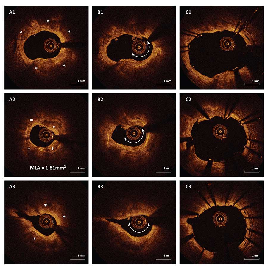
Figure 2. Matching OCT frames before orbital atherectomy (A1, A2, A3), after orbital atherectomy (B1, B2, B3) and after stenting (C1, C2, C3)
OCT Pre
OCT After OA
OCT After Stent
Ask the Experts

Ask the Experts
We are ready to help you.
ask now
ask now
Take Quiz

Take Quiz
Know about OCT? Test your knowledge.
try it now
try it now
Meet the Team

Meet the Team
Read about the team behind the apps.
learn more
learn more
Contact Us

Contact Us
Reach out to us with general comments or questions.
connect now
connect now