


Recurrent Spontaneous Coronary Artery Dissection
Operators: Annapoorna Kini, MD,
Samin Sharma, MD
Samin Sharma, MD
Interventional Fellow: Sunny Goel, MD
40-year-old female underwent PCI two months prior, one month post partum, due to chest pain and SOB. SCAD was detected and treated with two stents placed in the LM and mLAD. One week following previous PCI, patient experienced NSTEMI and was treated medically. The patient was transferred to MSH for SCAD management with peak troponin level 25 with decompensated HF.
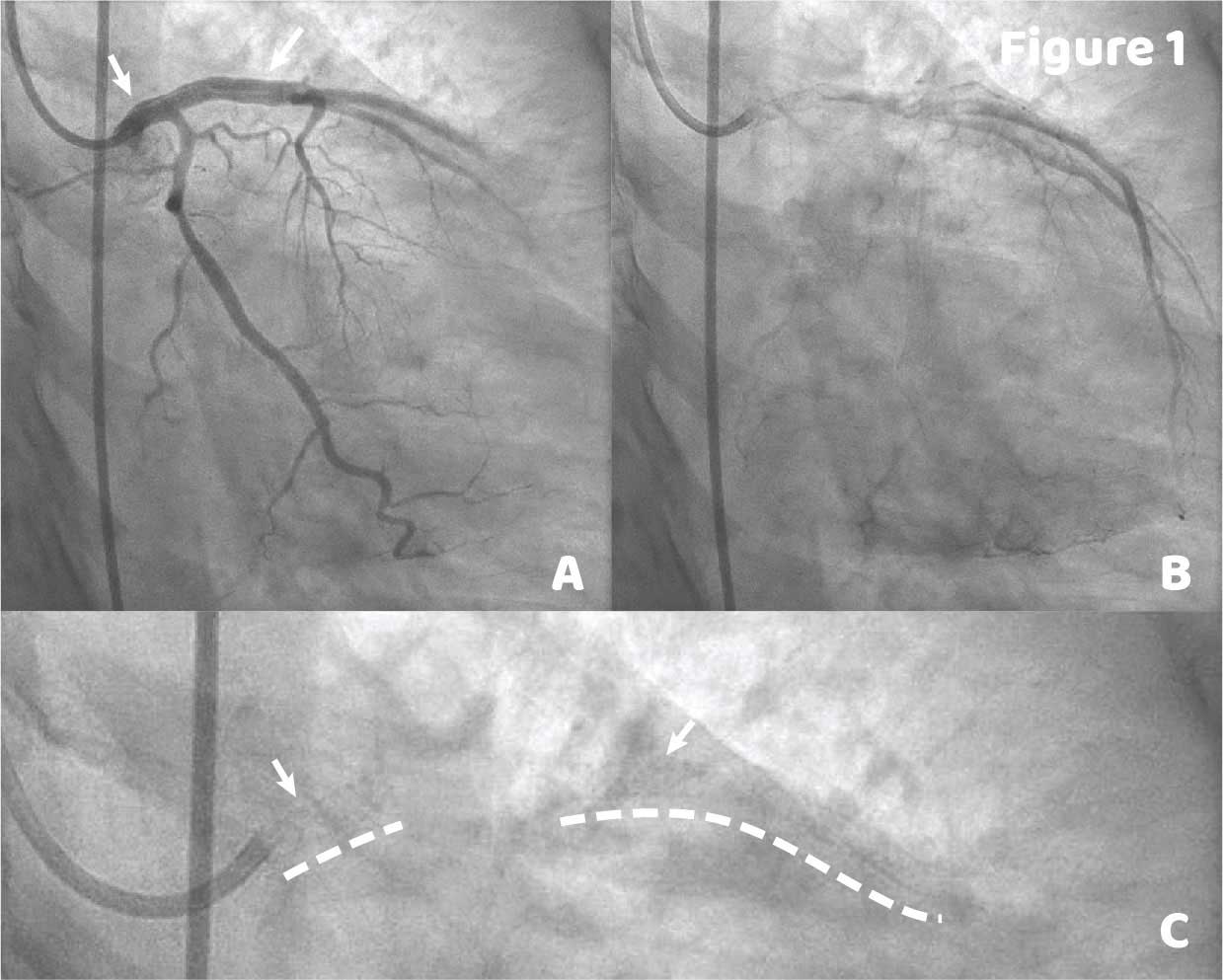
Coronary angiography shows the presense of a double lumen of the known SCAD from the prox LM stent to the proxial LAD stent (Figure 1A) resulting in TIMI 1 flow (Figure 1B). Figure C shows pre-injection frame with location of LM-pLAD and mLAD stents marked with dashed lines and arrows matching the false lumen arrows in Figure 1A.
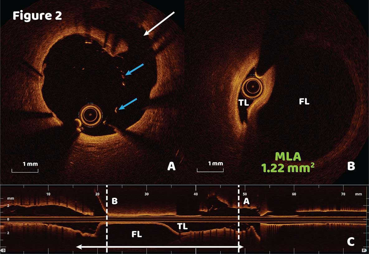
OCT reveals a 34 mm false lumen whose entry site is within the the proximal LM stent (Figure 2A, arrow) and terminates 5 mm past the proximal end of the mLAD stent. The MLA in the LAD was measured to be about 1.22 mm2 (Figure 2B). The full length of the SCAD is marked with a double-sided arrow in the longitudinal view of Figure 2C with the True Lumen (FL) and False Lumen (FL) marked. Proximal LM stent malapposition was detected (Figure 2A, blue arrows).
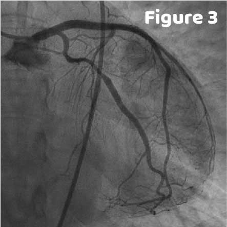
Based on OCT findings, a 4×23 mm DES was slected to slightly overlap the LM and LAD stents where the manjority of the false lumen was located. A 2×20 mm NC balloon was used for pre dilatation followed by a 4.5×12 mm NC balloon for post dilatation. Within the LM, dilatation with a 4.5×12 mm NC balloon was performed to improve apposition of the existing stent. Angiographic results below show good flow and that the false lumen was excluded (Figure 3).
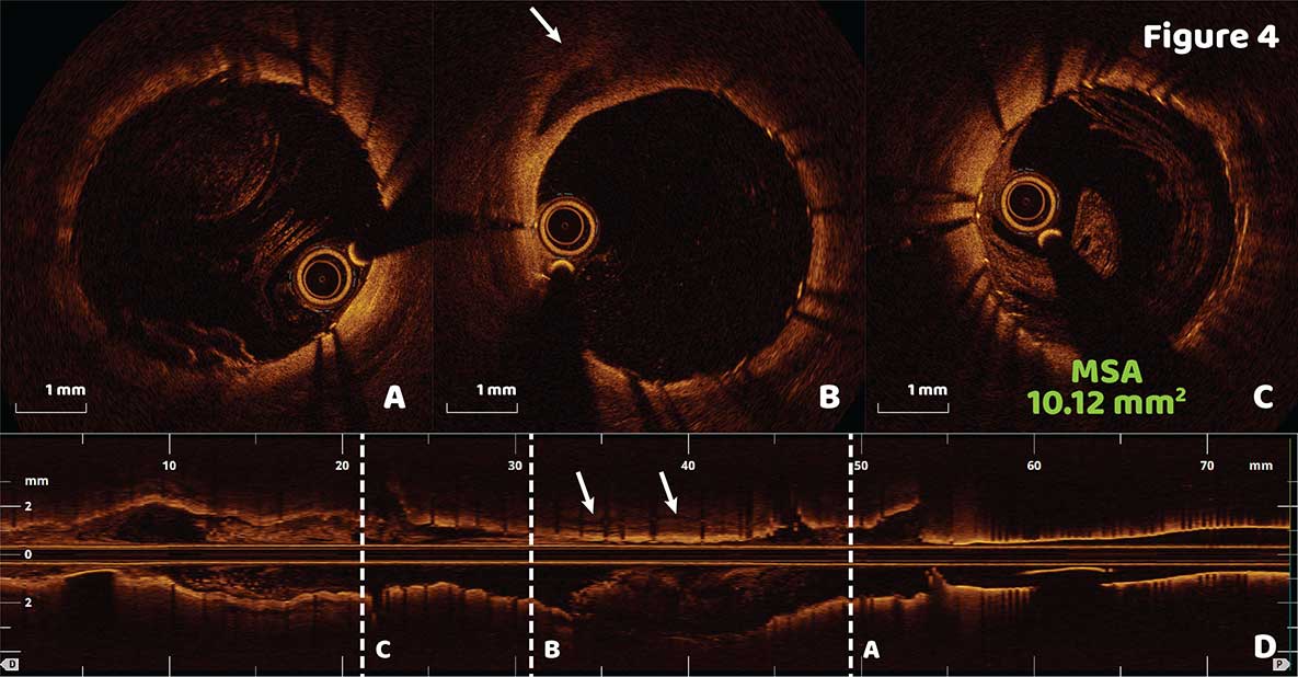
Post OCT findings confirmed the exclusion of the false lumen and significantly improved LM stent apposition (Figure 4A). The residual false lumen covered by the new stent is marked with arrows in Figure 4B and D. The LAD stent showed good expansion with a new MSA of 10.12 mm2 (Figure 4C).
Ask the Experts

Ask the Experts
We are ready to help you.
ask now
ask now
Take Quiz

Take Quiz
Know about OCT? Test your knowledge.
try it now
try it now
Meet the Team

Meet the Team
Read about the team behind the apps.
learn more
learn more
Contact Us

Contact Us
Reach out to us with general comments or questions.
connect now
connect now