


Principles Of Intracoronary Optical Coherence Tomography (OCT):
- Near-infrared light with a wavelength of approximately 1.3 micrometers generates high-resolution intra-coronary images of approximately 10-20 micrometers which is an order of magnitude higher than IVUS.
- Blood attenuates near infrared light therefore OCT requires clearing of blood from the lumen.
- Measures the echo time delay and intensity of the light reflected from internal structures within tissues to generate cross sectional images
- Light beam from OCT is separated into two arms: sample arm and reference arm.
- SAMPLE arm light travels to sample tissue then it is reflected, refracted or absorbed by the tissue and travels back to interferometer
- REFERENCE arm light is directed to a mirror which it reflects off of and goes to interferometer where it combines with the sample arm light and then is detected by the detector.
- Two types of OCT systems:
- TD-OCT (First Generation Time Domain) – reference arm is mechanically scanned by a moving mirror to produce time varying time delay
- FD-OCT (Second Generation Time Domain) – light source is swept therefore the interference of the light beam from the tissue and reference oscillates according to the frequency difference
- Limitation of TD-OCT is need for an occlusion balloon to remove blood from vessel for imaging.
- FD-OCT uses a fixed reference mirror and adjustable laser light source and can reach relatively higher imaging speeds.
Key Concepts:
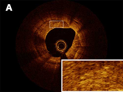
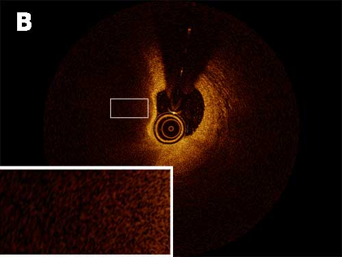
Attenuation is the reduction in the amplitude of a signal beyond the lumen. When there is a vessel with a lesion in the wall with low attenuation, it is easier to evaluate the lumen wall beyond the lesion whereas when there is high attenuation this evaluation becomes more challenging.
| Low Attenuation | High Attenuation |
|---|---|
| Calcific | Lipid |
| Fibrous | Red Thrombus |
| White Thrombus |
Calcific tissue has a low attenuation (Figure A)
Lipid is a highly attenuating tissue (Figure B)
Backscatter refers to signal intensity. The higher the backscattering, the brighter and more signal rich the image will appear. Low backscattering will appear dark (signal poor).
