M

Submit a Case

Complete the form below.
Please do not include any patient information.

  1. Home
  2. ComplicAID Case Reviews
  3. Burr Entrapment – Case 2

Burr Entrapment – Case 2

Clinical Presentation

  • 87-year-old male who presented with chest pain (CCS Class II).

Past Medical History

  • HTN, HLD, DM, CAD s/p PCI, Former Tobacco Use, Atrial Fibrillation
  • LVEF 68%

Clinical Variables

  • Stress MPI: Severe anterior ischemia.

Medications

  • Home Medications: Aspirin, Clopidogrel, Warfarin, Simvastatin, Metoprolol Tartrate, Diltiazem, Spironolactone, Allopurinol, Colchicine, Finasteride
  • Adjunct Pharmacotherapy: Clopidogrel, Bivalirudin

Pre-procedure EKG

Angiograms

Previous Next
Left coronary artery angiography
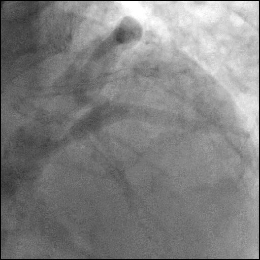
  • 90-95% calcified lesion in the ramus intermedius (RI).
1 of 15
1 of 15

Left coronary artery angiography

  • 90-95% calcified lesion in the ramus intermedius (RI).
2 of 15
2 of 15

Rotational atherectomy of the RI using a 1.5mm burr at 150k RPM.

3 of 15
3 of 15

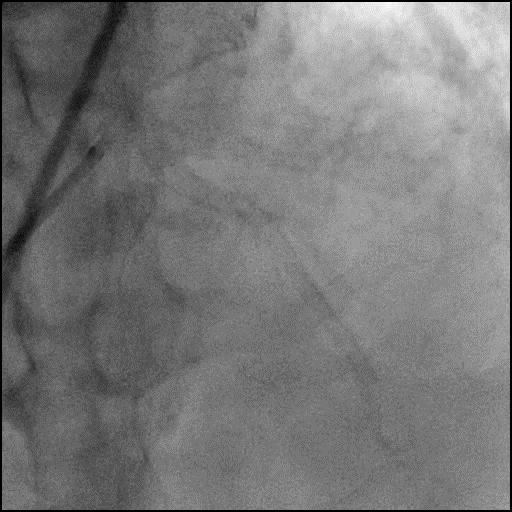
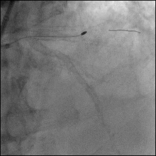
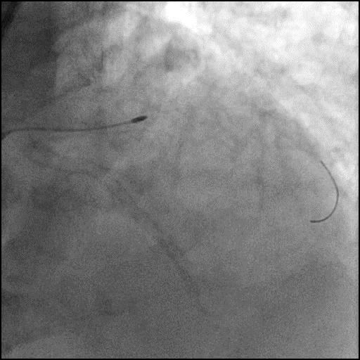
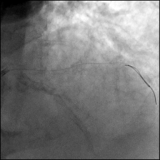
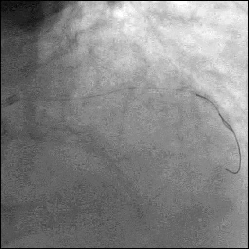
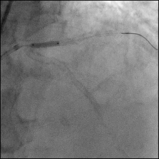
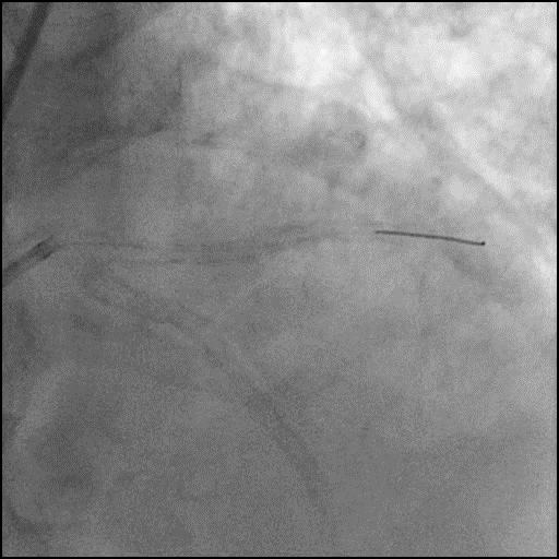
Rota burr stalled and became entrapped within the lumen of the RI.

4 of 15
4 of 15

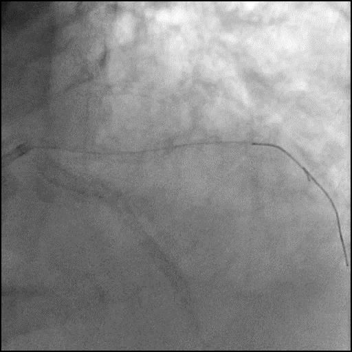
Mother (EBU 4.0/7 Fr) and child catheter (Guideliner 6 Fr) technique was used to retrieve the entrapped burr.

5 of 15
5 of 15

After successful retrieval of the 1.5mm burr, rotational atherectomy of the RI was repeated using a smaller 1.25mm burr at 150k RPM.

6 of 15
6 of 15

Angiography of the RI after rotational atherectomy.

7 of 15
7 of 15

Pre-dilatation of the RI lesion with a Quantum Apex 2.5/15mm balloon.

8 of 15
8 of 15

Angiography of the RI after lesion pre-dilatation.

9 of 15
9 of 15

Cutting balloon angioplasty of the RI lesion with an Angiosculpt 2.5/10mm balloon.

10 of 15
10 of 15

Angiography of the RI after cutting balloon angioplasty of the lesion was performed.

11 of 15
11 of 15

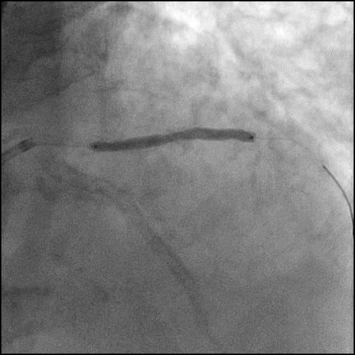
Deployment of a Resolute 3.0/38mm stent in the RI.

12 of 15
12 of 15

Angiography of the RI after stent placement.

13 of 15
13 of 15

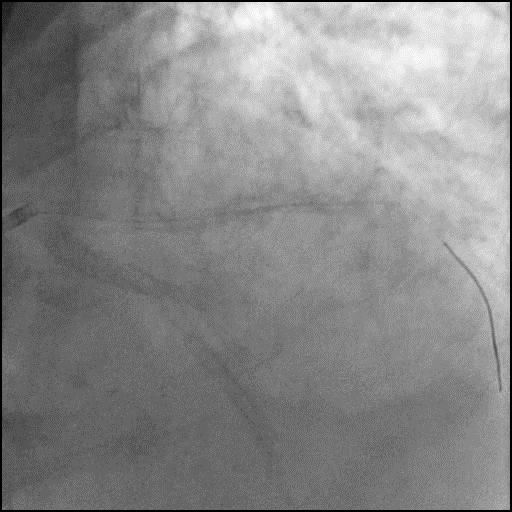
Deployment of a Resolute 3.0/12mm stent in the RI overlapping with the proximal stent edge of the previously placed stent.

14 of 15
14 of 15

Angiography after post-dilatation of the stent placed in the RI.

15 of 15
15 of 15

Final angiography showing successful intervention of the RI.

Post-procedure EKG

Case Overview

  • Underwent intervention of the RI.
  • Procedure was complicated by entrapment of a 1.5mm rota burr in a long calcified segment of the RI.
  • Mother and child catheter technique was used to successfully retrieve the entrapped burr.
  • Procedure was continued and successful intervention of RI was performed.
  • Troponin-I peaked at 0.1 ng/mL and CK-MB peaked at 2.8 ng/mL.
  • Patient was discharged home next day without further sequelae.

Learning Objectives

  • What is the likely explanation or reason why the complication occurred?
    • Two mechanisms for burr entrapment include:
      • ‘Kokesi’ phenomenon: When performing rotablation at high RPM, frictional heat is generated and it may enlarge the space between plaque. In addition, the coefficient of friction when the burr is in motion is less than that at rest, which may facilitate the burr to pass the calcified lesion easily without debulking a significant amount of calcified tissue. Once the burr traverses the lesion, and the plaque cools the between the plaque is again reduced, and the ledge of calcium proximal to the burr prevents the withdrawal of the burr, which is known as ‘Kokesi’ phenomenon, a name given after a Japanese doll.
      • Burr can become entrapped within a severely calcified ,long and/or angulated lesion when the burr is advanced aggressively. When a large burr is pushed forcefully against this kind of lesion without an appropriate pecking motion, significant decelerations occur and this produces more debris which increases the risk for slow flow/no-reflow and burr entrapment.
  • How could the complication have been prevented?
    • The burr is oval shaped and coated with diamonds at its distal end, allowing for antegrade ablation. However, the proximal end is smooth and not coated with diamonds, prohibiting retrograde ablation. If a burr is advanced beyond a tight calcified lesion or embedded in a long, angulated and heavily calcified lesion, it can be entrapped. Burr entrapment can often be avoided using the following techniques/strategies:
      • Use a gentle pecking motion with shorter runs of ablation (<20s).
        • Do not exert excessive forward force during burr advancement. If the burr is advanced aggressively, it causes decelerations and can become embedded in the calcified lesion.
        • The risk for burr entrapment is greater when the lesion is long and heavily calcified, and the vessel is highly angulated.
      • When advancing the burr, avoid decelerations >5000 RPM because this results in more debris production and increases risk for slow flow/no-reflow and burr entrapment.
      • When using a smaller burr, avoid using a higher speed of rotation (>180k RPM) to prevent ‘Kokesi’ phenomenon.; optimal speed is around 150k RPM.
      • If the vessel or lesion is highly tortuous/angulated, a stiffer wire can be used to straighten the vessel or lesion to lessen the resistance and reduce wire bias.
    • Avoid performing rotational or orbital atherectomy in vessels which are highly tortuous, especially if severe wire bias is present. Consider using Intravascular Lithotripsy (IVL) (off label use in the USA) for plaque modification and treatment of calcified CAD.
  • Is there an alternate strategy that could have been used to manage the complication?
    • Several bailout techniques can be used to retrieve a trapped burr, but prior to proceeding forward.
      • Assure patient is adequately anticoagulated (ACT >300) before attempting percutaneous retrieval.
      • Administer intracoronary vasodilators to facilitate antegrade coronary flow and relieve possible spasm.
    • Potential strategies for retrieval of an entrapped burr include the following:
      • 1st: Manual traction of the rotablator system by pulling the burr, guidewire and/or guide catheter as a unit. This can be performed on or off Dynaglide. The vessel is at risk for perforation, dissection, and abrupt vessel closure  (AVC). In addition, the burr shaft can fracture. If you are pulling the burr and guidewire as unit (and not the guide catheter), remember to disengage the guide catheter to prevent injury to the coronary artery from it deep seating during traction.
      • 2nd: Pass a second wire (hydrophilic-coated guidewire) beyond the trapped burr, followed by balloon dilatation around the burr. This may alter the architecture of the calcified lesion and possibly free the trapped burr. However, a 4.3 Fr rotablation drive shaft sheath may prohibit introduction of a balloon catheter into the guide catheter (consider this possibility if using a 6 or 7 Fr guide catheter). To overcome this, use a two-catheter strategy (Ping-Pong technique) where a second vascular access is obtained and equipment necessary for burr retrival is introduced through. If a single guide catheter strategy is preferred, there are two options. On approach includes cutting the rota system near the advancer, and remove the sheath to expose the driveshaft surrounding the rota-wire. This approach makes room for introduction of a second guidewire and balloon. This approach is useful when using a 6 Fr guide catheter. Alternatively, you can upsize the access sheath and guide catheter to a 8 Fr.
      • 3rd: Mother-child catheter technique can be used to wedge the burr and facilitate retrieval. The system is cut near the advancer, and the Teflon sheath is removed exposing the driveshaft which surrounds the rota-wire. A child catheter (monorail 5 Fr Guideliner or 5 Fr Guidezilla) is inserted over the exposed drive shaft and positioned as close as possible to the entrapped burr. With simultaneous traction on the burr shaft and counter-traction on the child catheter, the catheter tip wedges between the burr and the surrounding plaque, exerting a larger and direct pulling force to retrieve the burr.
      • 4th: Exclusion with a stent (As was done in this case).
      • 5th: Emergent surgical retrieval should always be the last option for removing an entrapped burr, but is often required.
  • What are the important learning points?
    • Interventional cardiologists who use rotablation, must be familiar with complications associated with its use and their management, particularly burr entrapment which is a rare but serious complication (incidence is ~0.4%, and occurs more frequently when rotablation is used off-label).
    • Use a burr which is appropriately sized for the vessel; ideally the burr should be 0.5-0.6x the reference vessel size when performing a plaque modification strategy and 0.8-0.85x the reference vessel size when performing a debulking strategy (STRATA trial). With plaque modification strategy the purpose is to disrupt the calcium so it allows for safe passage of devices and easier expansion of device, balloons, and stents. With debulking, the aim is to break the calcium into particles so it can move through the coronary artery and eventually washout of the coronary microcirculation.
    • Consider upfront use of an extension catheter to navigate across a tortuous vessel. With the extension catheter already in position a quick bailout strategy may be feasible should a complication arise while performing rotational atherectomy.
    • Prior to retracting the burr using the various techniques above, consider disengaging the guide catheter and holding it fixed with one hand (usually the left) to prevent injury to the coronary artery from it deep seating while pulling the equipment with the opposite hand (usually the right).
Educational Content

ROTATIONAL ATHERECTOMY COMPLICATIONS

  • Common complications associated with rotational atherectomy are:1
    • Slow flow / No-reflow
    • Coronary Dissection
    • Coronary Perforation
    • Burr entrapment

Slow flow / No-reflow

  • It is most feared and preventable operator-dependent complication of Rotational Atherectomy (RA)
  • Incidence- 2.6% in the drug-eluting stent era2
  • Omens of slow-flow / no-reflow include sudden decelerations and visual, tactile or auditory clues of high resistance to burr advancement
  • Be mindful of incident chest pain, ST-segment elevations, hemodynamic instability, and bradyarrhythmia while burring which could signal no-reflow phenomenon
  • Prevention
    • Optimal antiplatelet and anticoagulant therapy
    • Continuous flush cocktail
    • Smaller burr sizes (Max burr to artery ratio 0.4-0.6)
    • Lower speeds (140-150K rpm)
    • Short ablation runs of 15-20 seconds
    • Pause between runs
  • Treatment
    • Correction of hypotension with fluids, vasopressors, and pacing as required
    • Administration of intracoronary vasodilators, such as adenosine, nitrates, nitroprusside, nicardipine, and verapamil administered distally in the vessel
    • If hemodynamically unstable, insertion of an intraaortic balloon pump to augment coronary perfusion pressure

Coronary Dissection

  • Dissections during RA are described and graded in standard fashion using the NHLBI classification system (A-F)
  • Incidence: 1.7% - 5.9% in the drug-eluting stent era 3
  • Like slow-flow / no-reflow, dissection can present with signs and symptoms of acute myocardial ischemia including chest pain, ST-segment elevations, and hemodynamic or electrical instability
  • Prevention:
    • Avoid rotablation in excessively tortuous vessels
    • Avoid excessive angulation while burring
    • Smaller burr sizes
  • Treatment:
    • Stop further ablation
    • Maintain wire position
    • Expeditious completion of PCI via balloon angioplasty and stenting if feasible

Coronary Perforation

  • Perforation represents a more severe variant of dissection in which disruption extends through the full thickness of the arterial wall.
  • Incidence: 0-2% in the drug-eluting stent era3
  • Coronary perforations during RA are described and graded in standard fashion using the Elis classification scheme (I-III)
  • Although RA is considered a risk factor for perforation,4 the majority of type III perforations result from balloon angioplasty 5
  • Risk factors: lesion-specific predictors of perforation include eccentricity, tortuosity, length >10 mm, and location in the right coronary artery or left circumflex artery
  • Prevention:
    • Correct burr sizing
    • Avoid aggressive burring
    • Avoid excessive angulation
    • Lower speeds
  • Treatment:
    • Stop further ablation
    • Maintain wire position
    • Discontinuation of anticoagulation
    • Prolonged balloon inflation (10-15 min) proximal or at site of injury. If still bleeding, repeat prolonged balloon inflation
    • If extravasation persists, seal the site with either occlusive coils [perforation site distal main vessel] or by implantation of polytetrafluoroethylene (PFTE) covered stent [perforation siteproximal main vessel, distal side branch which can be excluded with covered stent]
    • If extravasation still persists or site of injury is proximal main vessel with bifurcation (covered stent not an option) consider emergent surgery
    • Aggressive treatment with intravenous fluids, atropine, vasopressors, mechanical circulatory support if hemodynamics deteriorate

Burr Entrapment

  • Entrapment consists of burr embedding in a severe stenosis, preventing both further burr advancement and retrieval
  • Presence of diamond chips on the front, but not the rear, of the burr abets an opportunity for the burr to lodge within a lesion and become entrapped.
  • Once stuck and stalled within a lesion, retrograde ablation is not possible and friction associated with retrograde motion cannot be orthogonally displaced.
  • During ablation, the operator should be attentive to potential warning signs, which may be visual (lack of smooth advancement under fluoroscopy), auditory (pitch changes with variation in resistance encountered by burr), or tactile (resistance in advancer knob or excessive driveshaft vibration)
  • Incidence: 0.5% to 1%3
  • Prevention:
    • Meticulous relief of system tension before RA
    • Gentle pecking motions
    • Short ablation runs
    • Avoid excessive tortuosity
    • Do not stop spinning within a lesion
  • Treatment:
    • Apply forceful pull on the Rota wire with guide disengaged taking advantage of the wire’s 0.014 inches spring tip
    • Administer high dose of vasodilators and aggressively pull the Rota burr
    • Manual traction with on-Dynaglide or off-Dynaglide rotation
    • If above measures fail, potential catheter-based solutions to facilitate burr retrieval include
      • Obtain second arterial access and advance Fielder wire and a small (1~1.25mm) balloon distally, inflate at the level of Rota burr, then aggressively pull the Rota burr
      • Advance Guide extension catheters on the Rota Burr

7Fr Guide extension
Cut the Rota burr shaft at the connection outside the body, then advance 7Fr guide extension on the shaft until the Rota burr and pull aggressively.

6Fr Guide extension catheter
  • Cut the Rota burr and aggressively pull the Teflon covering sheath.
  • once done, then advance 6Fr Guide extension on the shaft until the Rota burr and pull aggressively
    • Subintimal tracking and reentry with balloon dilatation adjacent to the entrapped burr6, 7


ORBITAL ATHERECTOMY COMPLICATIONS

Differential sanding of Orbital atherectomy (OA) permits healthy tissue to flex away from the crown during orbit and can be used with speed selection options for low speed (80,000 rpm), high speed (120,000 rpm), or GlideAssist (5000 rpm).

Common complications associated with rotational atherectomy are:

  • Slow flow / No-reflow
  • Coronary Dissection
  • Coronary Perforation

Slow flow / No-reflow

Incidence: 0.9% in Orbit II trial and 0.7% in real work registry analysis.8, 9

The unique mechanism of action, differential sanding, combined with an average particle size of debris of 2.04 μm – smaller than a red blood cell – may contribute to lower rates of no-reflow and transient heart block with orbital atherectomy.10

Prevention
  • Optimal anticoagulant and antiplatelet therapy
  • Continue ViperSlide infusion
  • Always keep the crown advancing or retracting with slow advancement (1mm/sec)
  • Short run timetime ( <20 seconds)
  • Rest time = or > run time
Treatment
  • Ensure to keep the optimal blood pressure (SBP > 100Hg) and give fluids, vasopressors, and pacing as needed
  • Administer intracoronary vasodilators, such as adenosine, nitrates, nitroprusside, nicardipine, and verapamil administered distally in the vessel if necessary, via twin-pass dual access catheter
  • If hemodynamically unstable, place an intra-aortic balloon pump to augment coronary perfusion pressure

Coronary Dissection

  • Coronary artery dissection can be categorized by using NHLBI classification system (A-F)
  • Incidence: 3.4 % in Orbit II trial and 0.9% in Real world registry8, 9
  • Dissection can manifest with acute onset of chest pain, new EKG changes with ST elevations, and hemodynamic or conduction disturbances.
Prevention
  • Avoid high speed run
  • Avoid in very tortuous coronary anatomy or > 2 bends exceeding 90° angulations
  • Use of ViperWire advance with flex tip in a setting of tortuous artery
Treatment
  • Stop ablation immediately
  • Reassess hemodynamic and patient status, then give vasopressor as needed
  • Completion of PCI with balloon angioplasty and stent placement if possible

Coronary Artery Perforation

  • Coronary perforation is the most serious complication that can occur with OA.
  • With unique mechanism with pulsatile forces in OA, it can result in more significant tissue modification while having a higher risk of deep dissections and perforation.
  • Incidence: 0.7-2%8, 9, 11
  • Coronary perforations during OA can be graded in standard fashion using the Elis classification scheme (I-III).

Risk factors: Very tortuosity, use of higher speed (120,000 rpm), presence of lipid rich plaque and smaller calcification arc (less than 2 quadrants in OCT/IVUS)12
Prevention:
  • Use of lower speed (80,000 rpm)
  • Avoid excessive angulation( >2 bends exceeding 90° angulations)
  • Careful advancement when evidence of wire wrinkling from tension buildup is present leading to vessel straightening
  • Avoid high speed if the vessel diameter is less than 3 mm
  • Advance the burr slowly with a speed of 1 mm per second

Treatment:
  • Stop further ablation immediately
  • Maintain wire position
  • Discontinuation of anticoagulation
  • Prolonged balloon inflation (10-15 min) proximal or at site of injury. If still bleeding, repeat prolonged balloon inflation
  • If extravasation persists, consider to use coils or covered stent
  • Reassess the perforation and patient status with angiogram
  • Be ready to do emergency pericardiocentesis if necessary
  • If extravasation remains present and/or site of injury is proximal main vessel with bifurcation (covered stent not an option), consider emergent surgery
  • Aggressive treatment with intravenous fluids, atropine, vasopressors, mechanical circulatory support if hemodynamics deteriorates


References

  1. Sharma SK, Tomey MI, Teirstein PS, et al. North American Expert Review of Rotational Atherectomy. Circ Cardiovasc Interv. 2019;12(5):e007448. doi:10.1161/CIRCINTERVENTIONS.118.007448
  2. Naito R, Sakakura K, Wada H, Funayama H, Sugawara Y, Kubo N, Ako J, Momomura S. Comparison of long-term clinical outcomes between sirolimus-eluting stents and paclitaxel-eluting stents following rotational atherectomy.Int Heart J.2012; 53:149–153
  3. Tomey MI, Kini AS, Sharma SK. Current status of rotational atherectomy.JACC Cardiovasc Interv.2014; 7:345–353. doi: 10.1016/j.jcin.2013.12.196
  4. Shimony A, Joseph L, Mottillo S, Eisenberg MJ. Coronary artery perforation during percutaneous coronary intervention: a systematic review and meta-analysis. Can J Cardiol 2011;27:843–50.
  5. Al-Lamee R., Ielasi A., Latib A., et al. (2011) Incidence, predictors, management, immediate and long-term outcomes following grade III coronary perforations. J Am Coll Cardiol 4:87–95
  6. Sulimov DS, Abdel-Wahab M, Toelg R, Kassner G, Geist V, Richardt G. Stuck rotablator: the nightmare of rotational atherectomy.EuroIntervention.2013; 9:251–258. doi: 10.4244/EIJV9I2A41
  7. Tanaka Y, Saito S. Successful retrieval of a firmly stuck rotablator burr by using a modified STAR technique. Catheter Cardiovasc Interv.2016; 87:749–756. doi: 10.1002/ccd.26342
  8. Chambers JW, Feldman RL, Himmelstein SI, et al. Pivotal trial to evaluate the safety and efficacy of the orbital atherectomy system in treating de novo, severely calcified coronary lesions (ORBIT II). JACC Cardiovasc Interv. 2014;7(5):510-518. doi:10.1016/j.jcin.2014.01.158
  9. Lee MS, Shlofmitz E, Kaplan B, Alexandru D, Meraj P, Shlofmitz R. Real-World Multicenter Registry of Patients with Severe Coronary Artery Calcification Undergoing Orbital Atherectomy. J Interv Cardiol. 2016;29(4):357-362. doi:10.1111/joic.12310
  10. Sotomi Y, Shlofmitz RA, Colombo A, et al. Patient selection and procedural considerations for coronary orbital atherectomy system. Interv Cardiol 2016;11:33
  11. Parikh K., Chandra P., Choksi N., et al: Safety and feasibility of orbital atherectomy for the treatment of calcified coronary lesions: the ORBIT I trial. Catheter Cardiovasc Interv 2013; 81: pp. 1134-1139
  12. Kini AS, Vengrenyuk Y, Pena J, et al. Optical coherence tomography assessment of the mechanistic effects of rotational and orbital atherectomy in severely calcified coronary lesions. Catheter Cardiovasc Interv. 2015;86(6):1024-1032. doi:10.1002/ccd.26000


x

Left Main DK Crush Video ID