Case 17: In-stent restenosis treated with RA and lithotripsy
Case Presentation
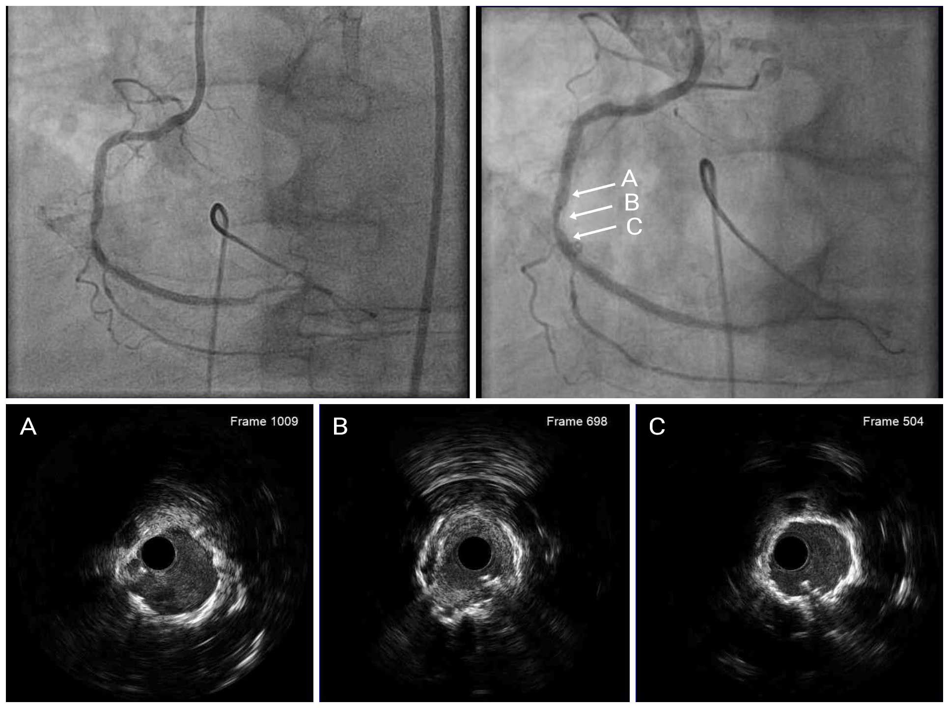
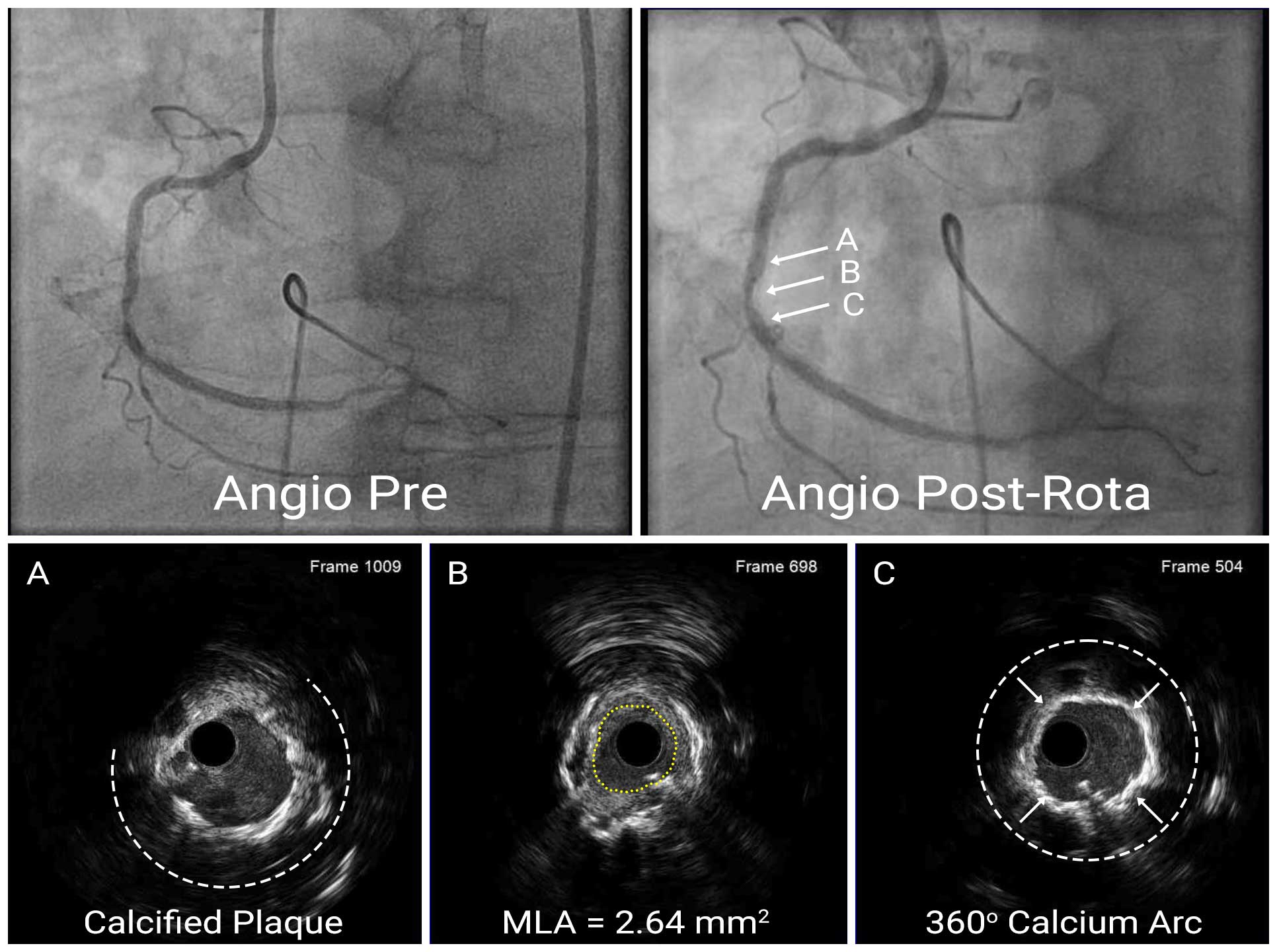
A 58-year-old male was admitted to our hospital for cardiac catheterization after a positive preoperative SPECT-MPI test for non-cardiac surgery. He has a history of hypertension, IDDM, ESRD (on dialysis), and has had multiple PCI procedures. Most recently, a year ago, DES were implanted mid RCA (3/38), distal RCA (2.5/24), and RPDA (2.5/28). Echocardiography showed good left ventricular systolic function (Ejection Fraction of 60%). A coronary angiogram showed 90-95% stenosis of the mid RCA. Since IVUS did not pass through the lesion, 1.5mm RA was performed first, followed by IVUS of the lesion. This showed an underexpanded stent, atherosclerosis in the stent, and circumferential calcium with a 360 degree arc outside the stent. The MLA of the lesion was 2.64mm².
IVUS After RA
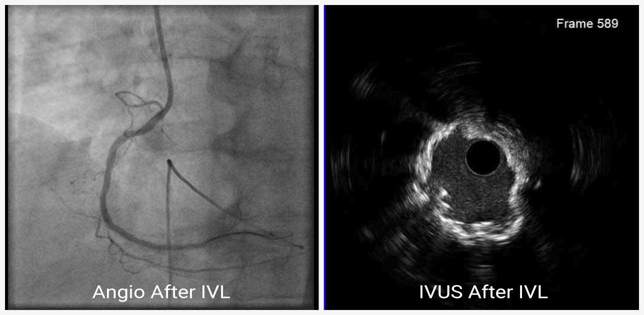
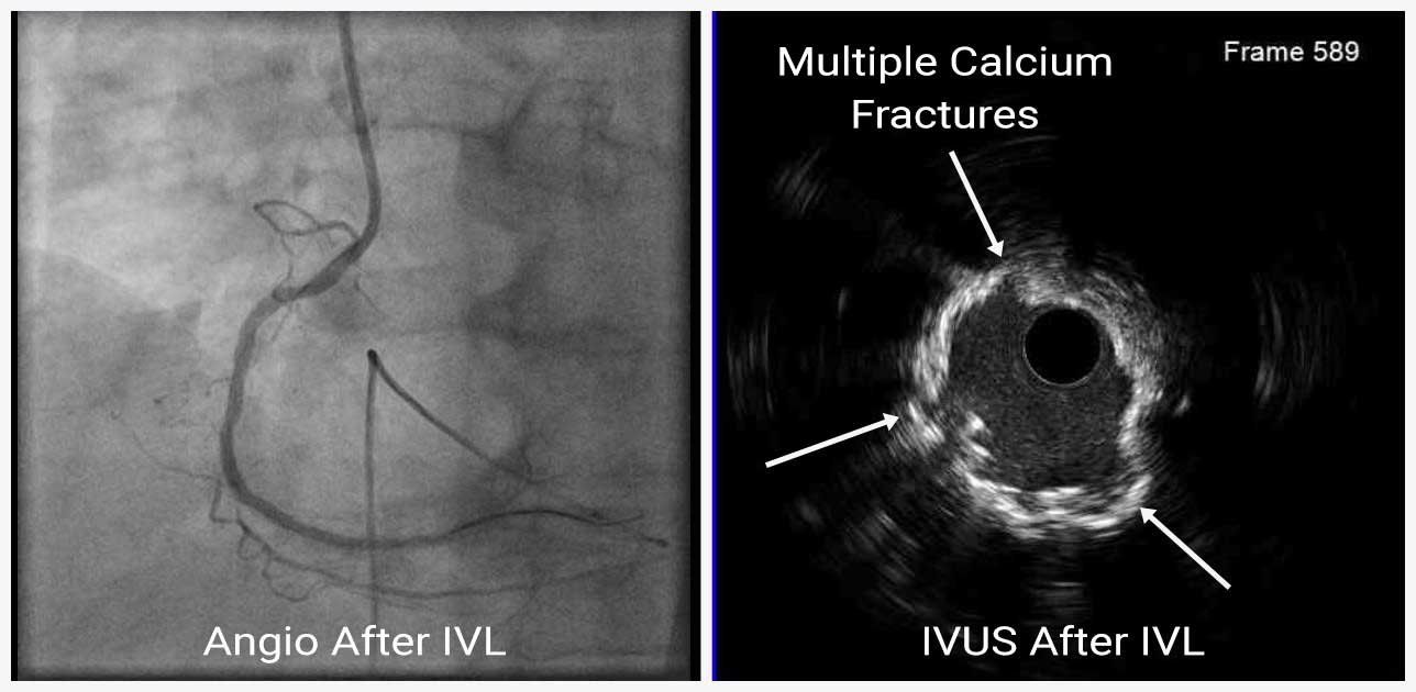
We planned for intravascular lithotripsy for calcified coronary segments and subsequent coronary stenting. The lesion was pre-dilated with 2.75mm x 15mm non-compliant (NC) balloon up to 20atm pressure. Then, the 3.0 mm x 12mm IVL balloon (Shockwave C2 IVL, Shockwave Medical) was placed across the mid RCA lesion, dilated up to 6atm pressure. Four balloon inflations with 10 shockwave pulse each were delivered to the lesion. Repeat IVUS images showed multiple fractures of calcified plaque at the lesion. The MLA was 4.66 mm² at the lesion after IVL.
IVUS After IVL
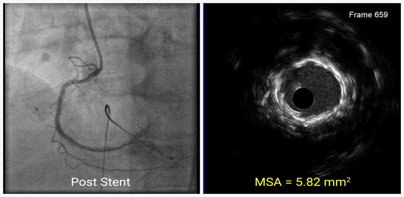
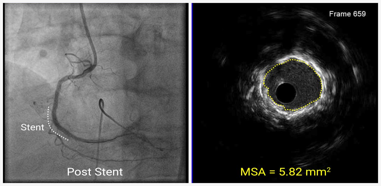
A 3.0mm x 23mm DES was deployed at the mid RCA and post dilated with a 3.0mm x 12mm NC balloon up to 20atm pressure. Repeat IVUS was performed following stenting. IVUS showed an improvement in MLA from pre angioplasty of 2.64mm² to post angioplasty MSA of 5.82mm².
IVUS After Stent
In cases with stents which have been previously implanted, it is often difficult to identify the presence or absence of calcification by angiography alone. In this case, IVUS could be used to identify the mechanism of restenosis, which may be due to severe calcification of the outside of the stent resulting in stent underexpansion and neointima inside the stent. Also, IVUS after IVL showed fractures of calcification in the lesion.


