Case 6: Calcified LAD/D1 bifurcation treated with mini-crush stenting
Case Presentation
A 74-year-old male presented with intermittent dizziness and left side chest pressure occurring anytime to outside hospital. He has a history of hypertension, hyperlipidemia, BPH and progressive aortic stenosis. His most recent echocardiography showed severe aortic stenosis (AVA0.84cm2), mild to moderate tricuspid regurgitation, good left ventricular systolic wall motion, with ejection fraction of 69%. He subsequently underwent coronary angiography at outside hospital which showed complex LAD lesion (severe lesion in the mid-portion-involving the large major D1-LAD 90%/D1 90%) and referred to our hospital for complex PCI.
Angio Pre
IVUS Pre LAD Pullback
IVUS Pre D1 Pullback
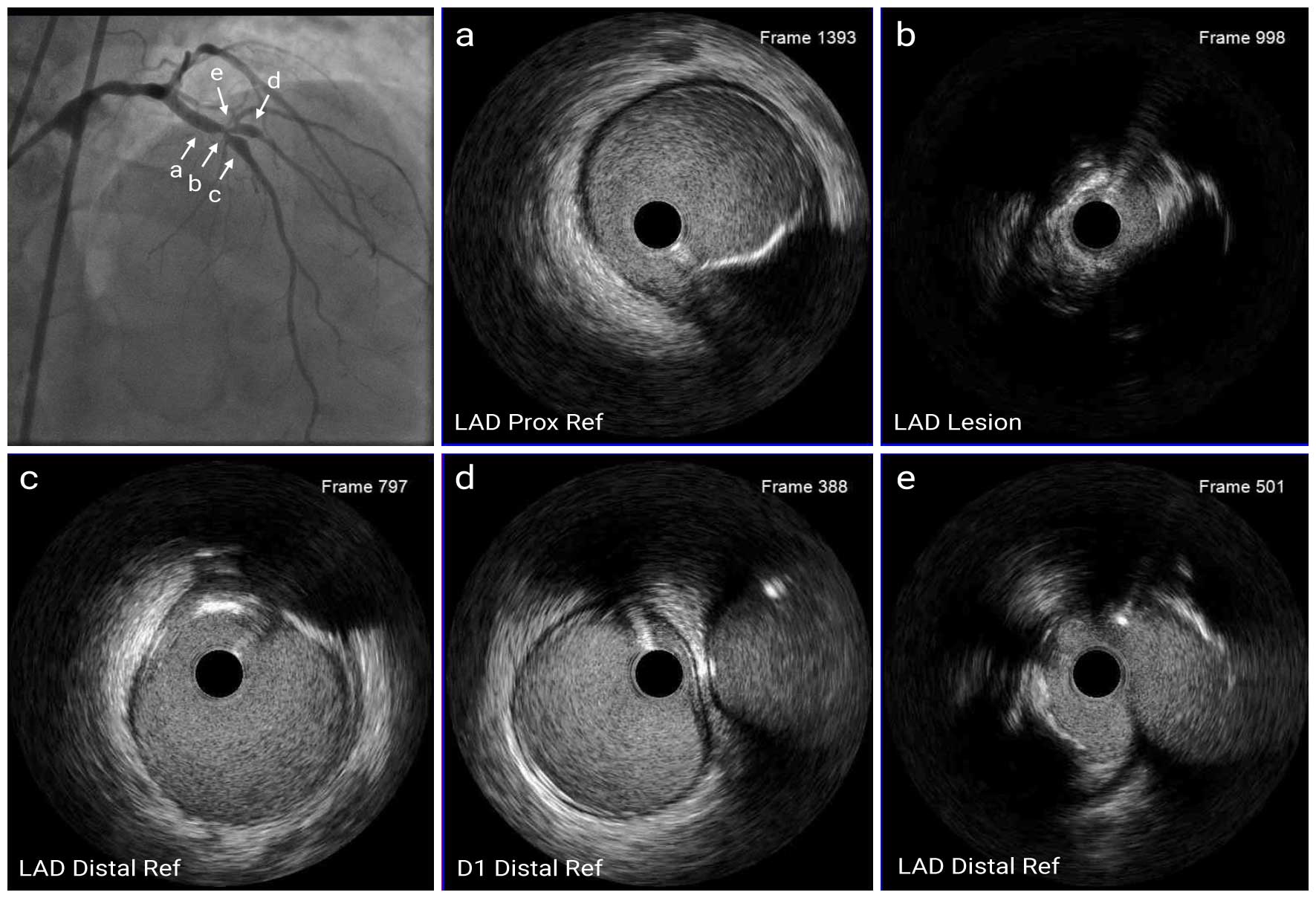
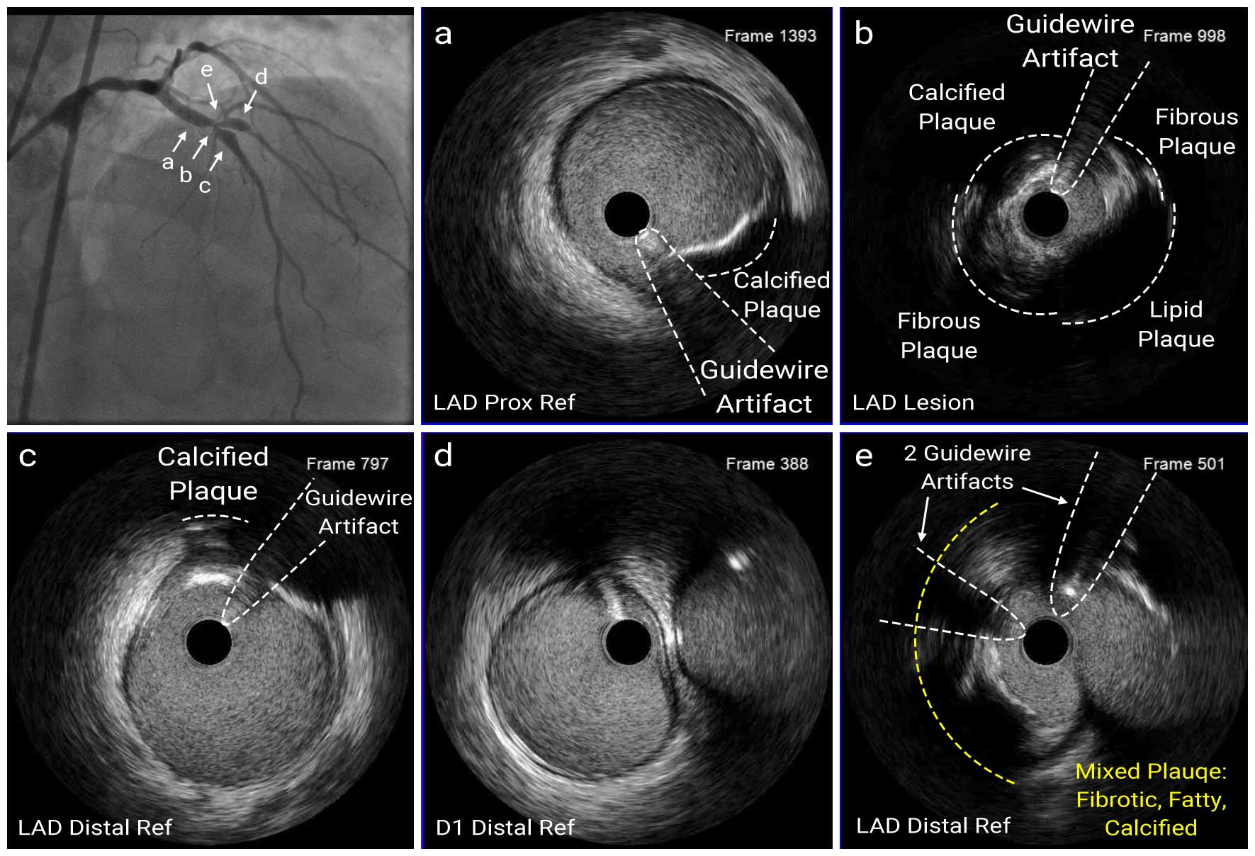
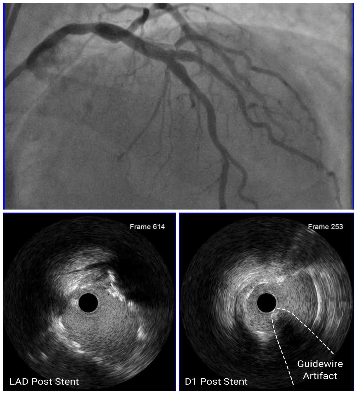
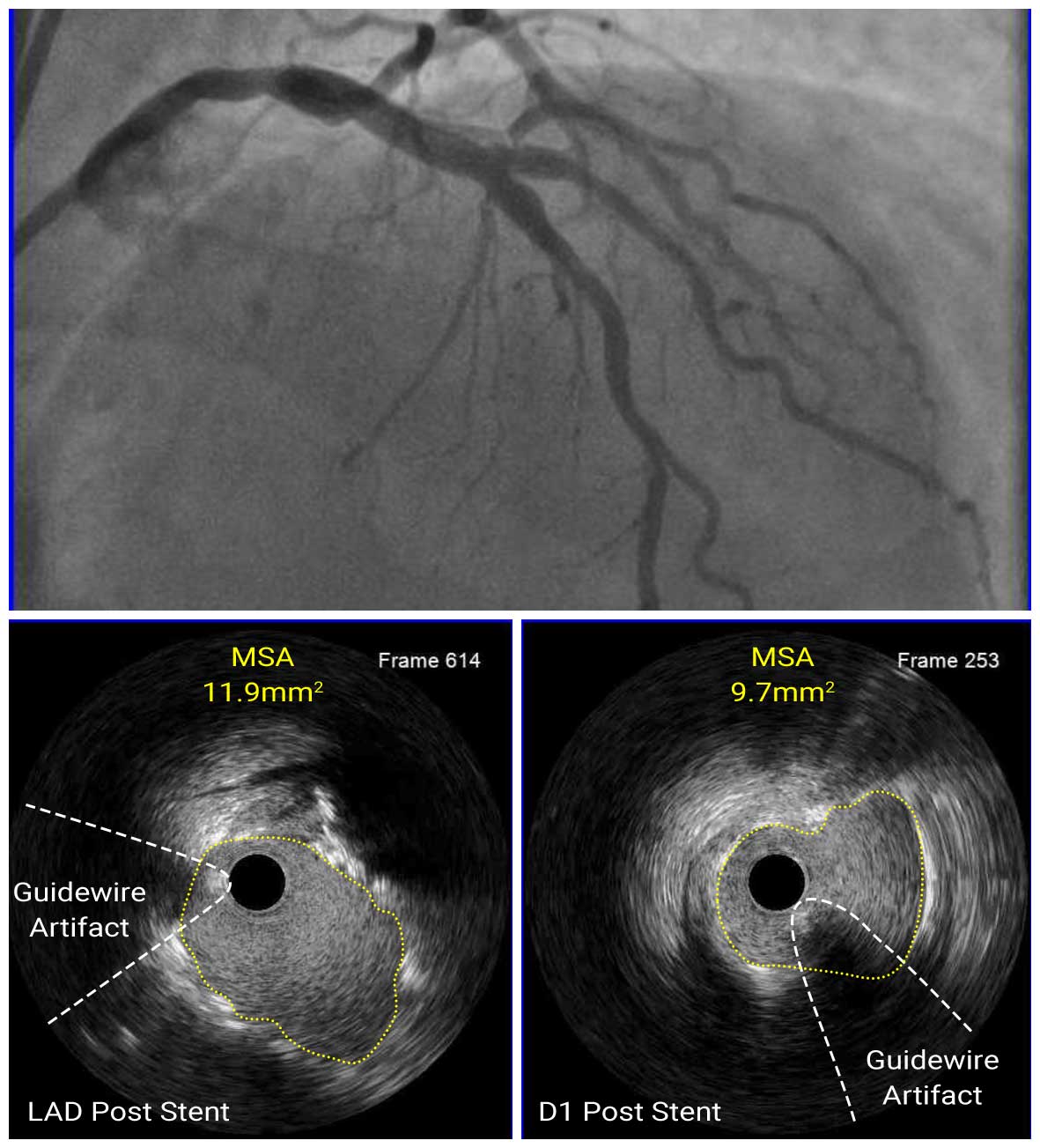
CAG showed (1,1,1) bifurcation lesion by medina classification, and IVUS images from LAD and D1 showed that the lesion was mainly composed of fibrous and lipid plaque with some shallow calcification, therefore we determined that atherectomy was not necessary. After lesion modification with 3.25mm cutting balloon dilated up to 16 atm pressure, mini crush technique was performed. 3.5mm x 8mm DES and 4.0mm x 12mm DES were deployed D1 and mid LAD, respectively. After stenting, IVUS showed that both stents were well expanded with the MSA 11.9mm2 and 9.7mm2 at LAD and D1, respectively, and there was no malapposition or dissection.
Angio Post
IVUS Post LAD
IVUS Post D1
In this case, angiography suggested the need for rotational atherectomy, but IVUS showed no severe calcification, therefore POBA was performed with cutting balloon and stent was well expanded. Imaging is useful in understanding the distribution of calcified lesions and in determining the treatment strategy.

