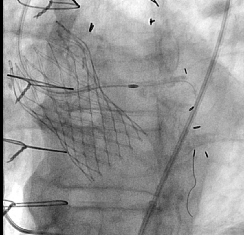
Dyspnea on Exertion
CoreValve
Case 3 – LM Rota/PCI
Case Presentation
- An 83-year-old male presented with dyspnea on exertion and leg swelling.
- Nuclear stress test showed moderate to severe lateral and posterolateral ischemia, mild inferior ischemia with partial scarring.
- Cardiac history: CAD (s/p CABG x 3 in 2006 : LIMA > LAD, SVG > D1 (occluded) jump to OM1, SVG > RPDA (patent), Severe AS s/p TAVR on 2/13/15, and CHB s/p PPM.
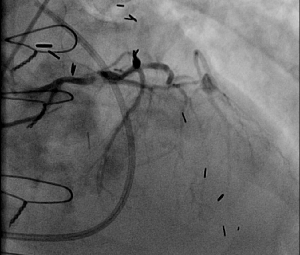
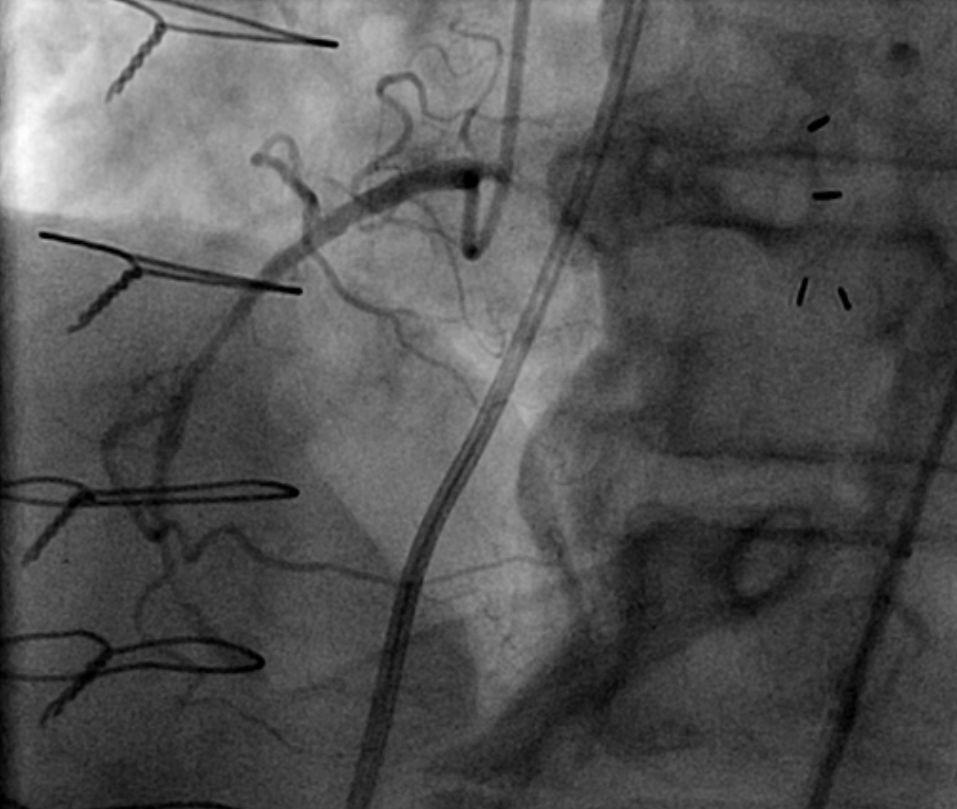
- Coronary angiogram revealed 3 vessels and left main disease with occluded SVG to LAD-D1-Jump OM1 and patent LIMA to mid LAD, SVG to RPDA.
- ECHO revealed LVEF of 65% and normal functioning bioprosthetic aortic valve with mild to moderate AR.
- With the presence of occluded vein graft to LAD-D1 and OM1, planned to perform PCI to left main disease.
A prior diagnostic angiogram was done by using JL4 for left coronary system and AR 2 for right system.
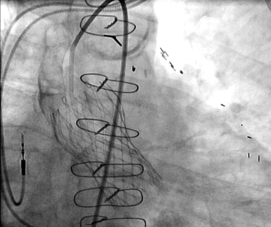
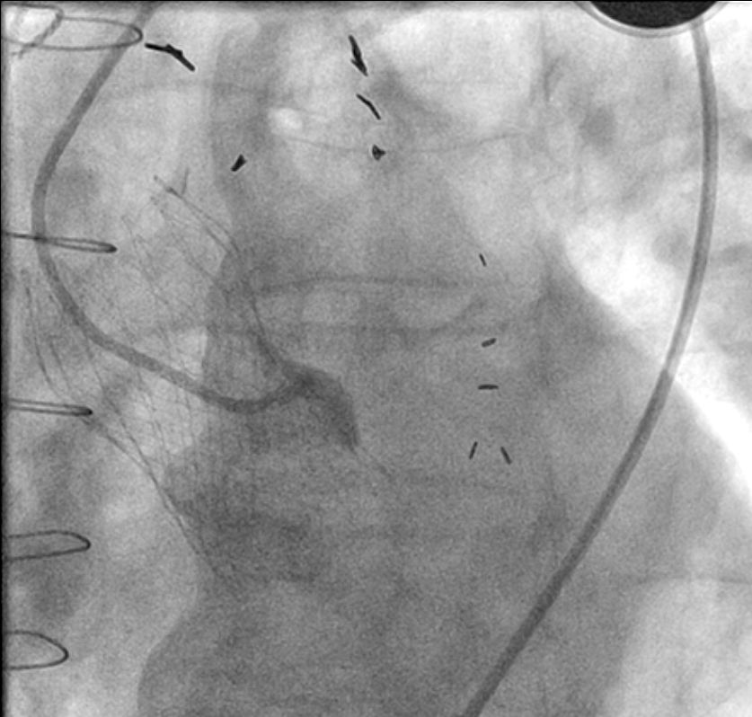
First, left coronary angiogram was attempted by VL 3.5 without success. Then, FL 3.5 guide catheter was used but the catheter was being directed into the inferior part of the left coronary system.
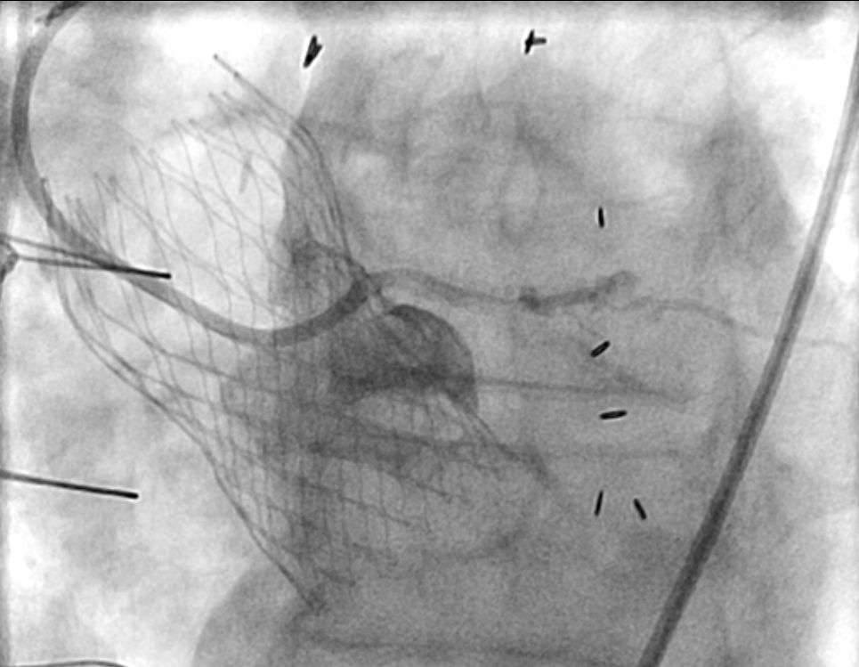
Then, we changed to FL 3 guide catheter and successfully engaged by railing the catheter over the J-wire.
As there was calcified severe LM disease, planned to do the lesion preparation using rotational atherectomy. Then, EBU 3.0 guide catheter was changed to have a better support for atherectomy (Rota 1.5 burr).
One DES (Promus 3.5/38) was placed in left main with an excellent result.
Learning Points
- Multiple guide catheters may sometimes require to perform coronary angiogram and intervention. This case had used VL 3.5 > FL 3.5> FL 3.0 > EBU 3.0.
- EBU 3.0 was used for a better support although FL 3.0 was successfully engaged.
- If necessary, selective coronary engagement can be done by railing the guide catheter over the J wire.

