


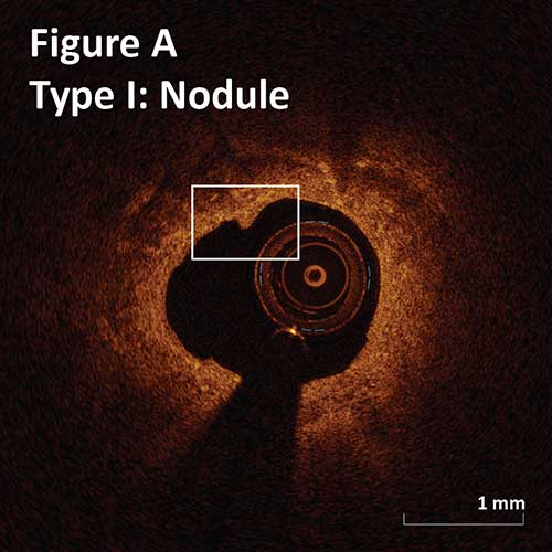
Type I. Superficial intimal flap with smooth luminal border:
Nodule: flaps length <0.5 mm. (Figure A)
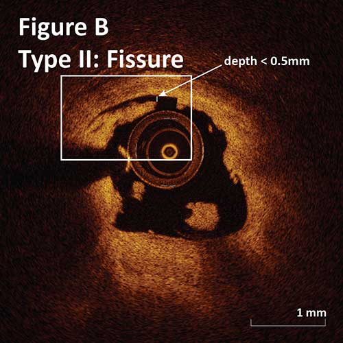
Type II. Deep intimal cut with irregular luminal border:
Fissure: 0–0.5 mm depth and 1 mm length. (Figure B)
Gutter: 0.5–1 mm depth and >1 mm length.
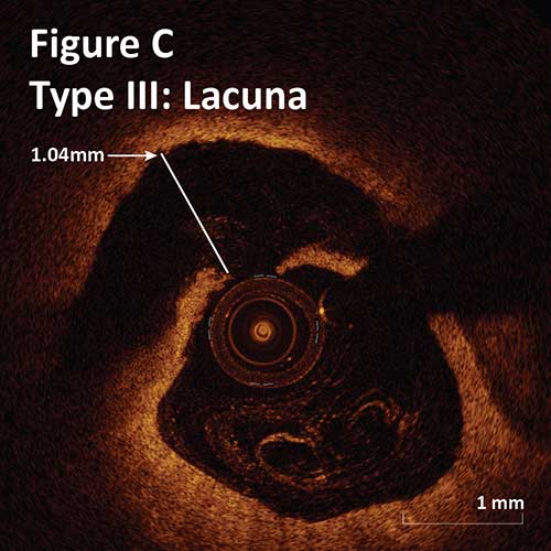
Type III. Deeper intimal-medial dissection with non-cylindrical lumen:
Crater: >1 mm depth and >3 mm length.
Lacuna: <1 mm depth and <3 mm length. (Figure C)
–There is no significant difference between the rate of each tissue modification seen with OA and RA. Craters and Lacunae occur in a third of lesions of OA and RA.1
–There is a difference between OA and RA regarding the severity of each tissue modification. Post OA lacunae were significantly deeper and have a bulging appearance.1
–Post OA modification of calcified plaque resulted in better stent apposition and expansion.1
–Overall, OA results in more severe tissue modification as compared to RA which may explain better stent placement but at the same time has higher risk of deep dissections, which may lead to perforation.1
1Catheter Cardiovasc Interv, 2015 Nov 15;86(6):1024-32.
Ask the Experts

Ask the Experts
ask now
Take Quiz

Take Quiz
try it now
Meet the Team

Meet the Team
learn more
Contact Us

Contact Us
connect now
