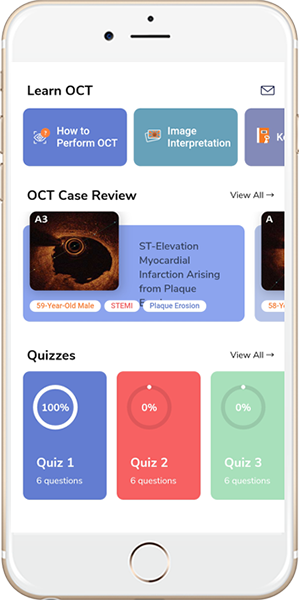
Image Interpretation, consensus standards and clinical use of OCT
- Huang D, Swanson EA, Lin CP et al. Optical coherence tomography. Science 1991;254:1178-81.
- Brezinski ME, Tearney GJ, Bouma BE et al. Optical coherence tomography for optical biopsy. Properties and demonstration of vascular pathology. Circulation 1996;93:1206-13.
- Jang IK, Bouma BE, Kang DH et al. Visualization of coronary atherosclerotic plaques in patients using optical coherence tomography: comparison with intravascular ultrasound. J Am Coll Cardiol 2002;39:604-9.
- Yamaguchi T, Terashima M, Akasaka T et al. Safety and feasibility of an intravascular optical coherence tomography image wire system in the clinical setting. Am J Cardiol 2008;101:562-7.
- Yabushita H, Bouma BE, Houser SL et al. Characterization of human atherosclerosis by optical coherence tomography. Circulation 2002;106:1640-5.
- Kume T, Akasaka T, Kawamoto T et al. Assessment of coronary arterial plaque by optical coherence tomography. Am J Cardiol 2006;97:1172-5.
- van Soest G, Regar E, Goderie TP et al. Pitfalls in plaque characterization by OCT: image artifacts in native coronary arteries. JACC Cardiovasc Imaging 2011;4:810-3.
- Tearney GJ, Regar E, Akasaka T et al. Consensus standards for acquisition, measurement, and reporting of intravascular optical coherence tomography studies: a report from the International Working Group for Intravascular Optical Coherence Tomography Standardization and Validation. J Am Coll Cardiol 2012;59:1058-72.
- Fedele S, Biondi-Zoccai G, Kwiatkowski P et al. Reproducibility of coronary optical coherence tomography for lumen and length measurements in humans. The American journal of cardiology 2012;110:1106-12.
- Kini AS, Vengrenyuk Y, Yoshimura T et al. Fibrous Cap Thickness by Optical Coherence Tomography In Vivo. J Am Coll Cardiol. 2017 Feb 14;69(6):644-657.
- Kini A, Narula J, Vengrenyuk Y, Sharma S. Atlas of Coronary Intravascular Optical Coherence Tomography. Springer 2017.
- Xing L, Higuma T, Wang Z et al. Clinical Significance of Lipid-Rich Plaque Detected by Optical Coherence Tomography: A 4-Year Follow-Up Study. J Am Coll Cardiol. 2017 May 23;69(20):2502-2513.
- Räber L, Mintz GS, Koskinas KC et al. Clinical use of intracoronary imaging. Part 1: guidance and optimization of coronary interventions. An expert consensus document of the European Association of Percutaneous Cardiovascular Interventions. Eur Heart J. 2018 Sep 14;39(35):3281-3300.
- Thomas W Johnson, Lorenz Räber, Carlo di Mario et al. Clinical use of intracoronary imaging. Part 2: acute coronary syndromes, ambiguous coronary angiography findings, and guiding interventional decision-making: an expert consensus document of the European Association of Percutaneous Cardiovascular Interventions. European Heart Journal, Volume 40, Issue 31, 14 August 2019, Pages 2566–2584.
Acute Coronary Syndrome:
- Tanaka A, Imanishi T, Kitabata H et al. Morphology of exertion-triggered plaque rupture in patients with acute coronary syndrome: an optical coherence tomography study. Circulation 2008;118:2368-73.
- Yonetsu T, Kakuta T, Lee T et al. In vivo critical fibrous cap thickness for rupture-prone coronary plaques assessed by optical coherence tomography. European Heart Journal 2011;32:1251-9.
- Jia H, Abtahian F, Aguirre AD et al. In vivo diagnosis of plaque erosion and calcified nodule in patients with acute coronary syndrome by intravascular optical coherence tomography. Journal of the American College of Cardiology 2013;62:1748-58.
- Prati F, Uemura S, Souteyrand G et al. OCT-based diagnosis and management of STEMI associated with intact fibrous cap. JACC Cardiovascular imaging 2013;6:283-7.
- Otsuka F, Joner M, Prati F, Virmani R, Narula J. Clinical classification of plaque morphology in coronary disease. Nature reviews Cardiology 2014;11:379-89.
- Saia F, Komukai K, Capodanno D et al. Eroded Versus Ruptured Plaques at the Culprit Site of STEMI: In Vivo Pathophysiological Features and Response to Primary PCI. JACC Cardiovascular imaging 2015;8:566-75.
- Higuma T, Soeda T, Abe N et al. A Combined Optical Coherence Tomography and Intravascular Ultrasound Study on Plaque Rupture, Plaque Erosion, and Calcified Nodule in Patients With ST-Segment Elevation Myocardial Infarction: Incidence, Morphologic Characteristics, and Outcomes After Percutaneous Coronary Intervention. JACC Cardiovascular interventions 2015.
- Kanwar SS, Stone GW, Singh M et al. Acute coronary syndromes without coronary plaque rupture. Nature reviews Cardiology 2016;13:257-65.
- Sugiyama T, Yamamoto E, Bryniarski K et al. Nonculprit Plaque Characteristics in Patients With Acute Coronary Syndrome Caused by Plaque Erosion vs Plaque Rupture A 3-Vessel Optical Coherence Tomography Study. JAMA Cardiol. 2018;3(3):207-214.
- Vergallo R, Porto I, D’Amario D et al. Coronary Atherosclerotic Phenotype and Plaque Healing in Patients With Recurrent Acute Coronary Syndromes Compared With Patients With Long-term Clinical Stability. An In Vivo Optical Coherence Tomography Study. JAMA Cardiol. 2019;4(4):321-329.
Stable Coronary Artery Disease:
- Kini AS, Yoshimura T, Vengrenyuk Y et al. Plaque Morphology Predictors of Side Branch Occlusion After Main Vessel Stenting in Coronary Bifurcation Lesions: Optical Coherence Tomography Imaging Study. JACC Cardiovasc Interv. 2016 Apr 25;9(8):862-865
- Kini AS, Vengrenyuk Y, Shameer K et al. Intracoronary Imaging, Cholesterol Efflux, and Transcriptomes After Intensive Statin Treatment: The YELLOW II Study. J Am Coll Cardiol. 2017 Feb 14;69(6):628-640
- Fujino A, Mintz GS, Matsumura M. A new optical coherence tomography-based calcium scoring system to predict stent underexpansion. EuroIntervention 2018;13:e2182-e2189 published online February 2018
- Okamoto N, Ueda H, Bhatheja S et al. Procedural and one-year outcomes of patients treated with orbital and rotational atherectomy with mechanistic insights from optical coherence tomography. EuroIntervention. 2019 Apr 20;14(17):1760-1767
- Okamoto N, Vengrenyuk Y, Bhatheja S et al. Stent Expansion and Endothelial Shear Stress in Bifurcation Lesions. Circ Cardiovasc Interv. 2019 Jun;12(6):e007911
Post Stent Evaluation and In-stent Restenosis:
- Kang SJ, Mintz GS, Akasaka T et al. Optical coherence tomographic analysis of in-stent neoatherosclerosis after drug-eluting stent implantation. Circulation 2011;123:2954-63.
- Ali ZA, Roleder T, Narula J et al. Increased thin-cap neoatheroma and periprocedural myocardial infarction in drug-eluting stent restenosis: multimodality intravascular imaging of drug-eluting and bare-metal stents. Circulation Cardiovascular interventions 2013;6:507-17.
- Parodi G, La Manna A, Di Vito L et al. Stent-related defects in patients presenting with stent thrombosis: differences at optical coherence tomography between subacute and late/very late thrombosis in the Mechanism Of Stent Thrombosis (MOST) study. EuroIntervention 2013;9:936-44.
- Karanasos A, Simsek C, Gnanadesigan M et al. OCT assessment of the long-term vascular healing response 5 years after everolimus-eluting bioresorbable vascular scaffold. J Am Coll Cardiol 2014;64:2343-56.
- Prati F, Romagnoli E, Burzotta F et al. Clinical Impact of OCT Findings During PCI: The CLI-OPCI II Study. JACC Cardiovascular imaging 2015;8:1297-305.
- Bastante T, Rivero F, Cuesta J, Alfonso F. Calcified Neoatherosclerosis Causing “Undilatable” In-Stent Restenosis: Insights of Optical Coherence Tomography and Role of Rotational Atherectomy. JACC Cardiovascular interventions 2015;8:2039-40.
- Maehara A, Ben-Yehuda O, Ali Z et al. Comparison of Stent Expansion Guided by Optical Coherence Tomography Versus Intravascular Ultrasound: The ILUMIEN II Study (Observational Study of Optical Coherence Tomography [OCT] in Patients Undergoing Fractional Flow Reserve [FFR] and Percutaneous Coronary Intervention). JACC Cardiovascular interventions 2015;8:1704-14.
- Ali ZA, Genereux P, Shlofmitz RA et al. Optical coherence tomography compared with intravascular ultrasound and with angiography to guide coronary stent implantation (ILLUMIEN III: OPTIMIZE PCI): a randomized control trial. Lancet 2016; 388: 2618–28.
We use cookies to ensure that we give you the best experience on our website. If you continue to use this site we will assume that you are happy with it.OKNoPrivacy Policy

